Meta-review: trends and predictions
A curated, semi-automated review of thousands of bleb papers: how they were found, read and counted, what the counts show, and what past and present reviews expected of the field.
This page is a meta-review: a review of the bleb literature as a whole rather than of any single question. Searches of databases and of the older literature, followed by sorting under written rules that we tested by hand, produced nine curated paper lists holding 5,175 dated papers from 1856 to 2026, and structured reading notes cover about 5,400 publications. We use this material to count how bleb research has grown and shifted, and to compare what reviews expected of the field with what happened. Counts measure activity, not importance; which work changed ideas is the subject of A long history of blebbing.
A map of the field
The map links the main ideas on this site: the molecules that set the mechanics of the cell surface, the forces and materials involved, the life of a single bleb, the kinds of blebs, and the cells and conditions in which they appear. Each arrow states a relation that the linked pages describe and cite; dashed lines join related ideas without implying cause.
Swipe sideways to see the whole map.
Hover over or tap a concept to see what it connects to; select it to read its definition and find the pages that treat it. The buttons hide or show each kind of concept.
Rho–ROCK Molecules
The small GTPase RhoA and the kinase it activates, ROCK (Rho kinase). ROCK raises myosin II activity by phosphorylating the myosin light chain and inhibiting its phosphatase; this is the best-studied signaling route to the contractility that drives blebs (Mills et al. 1998; Sahai & Marshall 2003).
Read more: Physics of blebs · Blebs in dying and injured cells · Glossary: Rho–ROCK
Rho–ROCK → myosin II: activates
myosin II Molecules
The motor protein that pulls on actin filaments and so contracts the cortex. Its activity, switched on by phosphorylation of its light chain through ROCK or MLCK, generates the cortical tension and pressure that drive blebs (Mills et al. 1998; Charras & Paluch 2008).
Read more: Physics of blebs · Glossary: myosin II
Milestones: M06, M08, M11, M12, M18
Rho–ROCK → myosin II: activates
blebbistatin → myosin II: inhibits
myosin II → actin cortex: contracts
blebbistatin Tools and theory
A small-molecule inhibitor of non-muscle myosin II, named because it stops blebbing (Anonymous 2002). It is widely used to test whether a bleb-driven process needs myosin activity (e.g. Liu et al. 2015).
Read more: Physics of blebs · Glossary: blebbistatin
Milestones: M12, M15, M18, M20
blebbistatin → myosin II: inhibits
actin turnover Molecules
The continual exchange of actin between the filaments of the cortex and the pool of free actin in the cytoplasm, through which the cortex renews itself within seconds to tens of seconds even when it looks unchanged (Fritzsche et al. 2013; Chugh et al. 2018). It is usually measured by bleaching fluorescent actin in a small area and timing the recovery of fluorescence (FRAP), and given as a half-time or a rate.
Read more: Physics of blebs · Blebs by the numbers · Glossary: actin turnover
actin turnover → actin cortex: renews
actin cortex Forces and materials
The thin meshwork of actin filaments, myosin II motors and cross-linking proteins that lies just under the plasma membrane, a fraction of a micrometer thick (M13). Its contraction sets the tension of the cell surface, and blebs form where it comes away from the membrane or breaks (Charras & Paluch 2008; Tinevez et al. 2009).
Read more: Physics of blebs · Glossary: cortex
Milestones: M02, M04, M06, M08, M10, M12, M13
myosin II → actin cortex: contracts
actin turnover → actin cortex: renews
actin cortex → cortical tension: generates
cortical tension Forces and materials
The tension within the plane of the cortex, generated mainly by myosin II pulling on actin filaments. Together with membrane tension it sets the pressure inside the cell that drives blebs (Tinevez et al. 2009).
Read more: Physics of blebs · Blebs by the numbers · Glossary: cortical tension
actin cortex → cortical tension: generates
cortical tension → intracellular pressure: raises
Laplace's law – cortical tension: relates
Laplace's law Tools and theory
The relation between the pressure difference across a curved surface, its tension and its radius of curvature: the smaller the radius, the larger the pressure difference a given tension holds. For membranes it is written T = ΔP·r/2, with T the tension, ΔP the pressure difference and r the radius (Sokabe & Sachs 1990); bleb studies use it to relate cortical tension, intracellular pressure and bleb size (Prothero & Spencer 1968; Tinevez et al. 2009).
Read more: Physics of blebs · Glossary: Laplace's law
Laplace's law – cortical tension: relates
Laplace's law – intracellular pressure: relates
intracellular pressure Forces and materials
The amount by which the hydrostatic pressure inside a cell exceeds the pressure outside. It is generated by the contracting cortex and inflates blebs; Laplace's law relates it to cortical tension and curvature (Charras et al. 2008; Tinevez et al. 2009).
Read more: Physics of blebs · Blebs by the numbers · Glossary: intracellular pressure
Milestones: M04, M05, M07, M12, M14
cortical tension → intracellular pressure: raises
Laplace's law – intracellular pressure: relates
poroelasticity → intracellular pressure: delays its spread
intracellular pressure → expansion: drives
intracellular pressure → initiation: detachment or rupture: drives
intracellular pressure → inverse bleb: if the outside pressure is higher
poroelasticity Tools and theory
A description of the cytoplasm as an elastic, porous solid mesh soaked in fluid. Because a local change in pressure spreads only as fluid flows through the mesh, pressure takes time to even out across the cell (Charras et al. 2005; Moeendarbary et al. 2013).
Read more: Physics of blebs · Glossary: poroelasticity
Milestones: M12
poroelasticity → intracellular pressure: delays its spread
ERM proteins Molecules
Ezrin, radixin and moesin, related proteins that, once activated by phosphorylation, bind both the plasma membrane and actin and so tie the membrane to the cortex. Their activity sets membrane–cortex attachment, and ezrin is among the first proteins to reach a retracting bleb (Charras et al. 2006; Diz-Muñoz et al. 2010).
Read more: Physics of blebs · Glossary: ERM proteins
ERM proteins → membrane–cortex attachment: provide
membrane–cortex attachment Forces and materials
The binding of the plasma membrane to the cortex beneath it, through linker proteins such as the ERM proteins. Measured as an energy per unit area by pulling membrane tethers, it opposes bleb formation: in zebrafish cells, weaker attachment gave more and larger blebs (Diz-Muñoz et al. 2010; Bergert et al. 2021).
Read more: Physics of blebs · Blebs by the numbers · Glossary: membrane–cortex attachment
ERM proteins → membrane–cortex attachment: provide
membrane–cortex attachment → initiation: detachment or rupture: opposes
reconstitution Tools and theory
Rebuilding a cellular process outside the cell from isolated or purified components. For blebs, actin–myosin networks are attached to the inner face of cell-sized lipid vesicles (liposomes), which bleb when the networks contract (Loiseau et al. 2016; Sakamoto & Murrell 2024; Miyazaki et al. 2026).
Read more: Physics of blebs · Glossary: reconstitution
reconstitution → initiation: detachment or rupture: reproduces both routes
inverse bleb Kinds of blebs
A bleb that grows into the cell instead of out of it. Where the pressure outside (of blood or intercellular fluid) is higher than inside, the membrane bulges inwards without actin and is then pulled back by actomyosin (Gebala et al. 2016; Schliffka et al. 2024).
Read more: Kinds of blebs · Physics of blebs · Glossary: inverse bleb
Milestones: M14
intracellular pressure → inverse bleb: if the outside pressure is higher
bleb The central concept
A rounded bulge of the plasma membrane that forms where the membrane comes away from the actin cortex beneath it, or where the cortex breaks. Pressure inside the cell, generated by contraction of the cortex, inflates it; a new cortex then assembles under the bulge, which usually retracts (Charras & Paluch 2008; Tinevez et al. 2009).
Read more: Blebology · Kinds of blebs · Glossary: bleb
Milestones: M03, M07, M08, M10, M12, M15, M19, M20
bleb → initiation: detachment or rupture: starts by
bleb → apoptotic bleb: in dying cells
bleb → large oncosome: said to pinch off as
bleb – nuclear bleb: not the same: nuclear envelope
initiation: detachment or rupture Life of a bleb
The two proposed ways a bleb can start: the membrane comes away from an intact cortex (detachment), or the cortex itself tears (rupture) (Tinevez et al. 2009). Both routes have been reproduced in liposomes, where actin–membrane coupling and cross-linking decide which one occurs (Miyazaki et al. 2026).
Read more: Physics of blebs · Glossary: rupture vs detachment
Milestones: M04, M08, M10, M12, M15
membrane–cortex attachment → initiation: detachment or rupture: opposes
intracellular pressure → initiation: detachment or rupture: drives
reconstitution → initiation: detachment or rupture: reproduces both routes
initiation: detachment or rupture → expansion: then
initiation: detachment or rupture → cortex scar: leaves the old cortex
bleb → initiation: detachment or rupture: starts by
expansion Life of a bleb
The life of a single bleb: it starts by membrane–cortex detachment or cortex rupture, grows as cytoplasm flows in, stops growing as a new cortex assembles under its membrane, and retracts as myosin contracts that cortex (Charras et al. 2006; Tinevez et al. 2009). A transient bleb completes the cycle in about a minute (García-Arcos et al. 2024, Trends Cell Biol ◆).
Read more: Physics of blebs · Glossary: bleb cycle
Milestones: M04, M07, M10, M12, M13
intracellular pressure → expansion: drives
initiation: detachment or rupture → expansion: then
expansion → new cortex assembles: stops when
new cortex assembles Life of a bleb
The building of new cortex on a patch of plasma membrane, where actin and its partner proteins are added faster than they are lost. Under a growing bleb, the bare membrane gains a new cortex in a set order in the cells studied, linkers such as ezrin first, then actin with early cross-linkers, then myosin II; this stops the bleb's growth and lets it retract (Charras et al. 2006; M13).
Read more: Physics of blebs · Glossary: cortex assembly
expansion → new cortex assembles: stops when
new cortex assembles → retraction: then
new cortex assembles → circus bleb: re-forms on one side only (proposed)
retraction Life of a bleb
The last step of the bleb cycle: myosin II contracts the new cortex under the bleb membrane and pulls the bleb back into the cell.
Read more: Physics of blebs · Kinds of blebs
new cortex assembles → retraction: then
retraction → transient bleb: a full cycle
cortex scar Life of a bleb
The old cortex left behind where a bleb starts: as the membrane moves away, the original cortex stays at the bleb base and disassembles over seconds to tens of seconds while a new cortex forms under the bleb membrane (Zatulovskiy et al. 2014; Asante-Asamani et al. 2024). Each transient bleb leaves one (García-Arcos et al. 2024, Trends Cell Biol ◆), and a scar is a common sign that a protrusion is a bleb rather than an actin-driven pseudopod.
Read more: Kinds of blebs · Glossary: cortex scar
initiation: detachment or rupture → cortex scar: leaves the old cortex
cortical flow Forces and materials
Movement of the actomyosin cortex along the cell surface, parallel to the membrane, toward regions where it contracts more strongly. In migrating cells it runs opposite to the direction of movement and is also called retrograde flow; in stable-bleb cells this rearward flow, coupled to the surroundings by friction, propels the cell (Liu et al. 2015; Ruprecht et al. 2015; Bergert et al. 2015).
Read more: Physics of blebs · Blebs in cell migration · Glossary: cortical flow
cortical flow → stable bleb: keeps the tip free of cortex
percolation → cortical flow: organizes (proposed)
percolation Tools and theory
In network physics, the point at which enough links have formed for a cross-linked network to span the whole system and transmit force across it. It has been applied to purified actomyosin gels (Alvarado et al. 2013) and to the cortex of stable blebs (García-Arcos et al. 2024, Dev Cell ◆); an older sense is fluid seeping through a porous gel (Cunningham 1995).
Read more: Physics of blebs · Glossary: percolation
percolation → cortical flow: organizes (proposed)
cytokineplast Cells, health and disease
A motile piece of a cell without a nucleus that has broken away carrying the machinery for movement. The word was coined for the heat-released leading edge of neutrophils (Malawista & de Boisfleury Chevance 1982) and is now also used for detached stable blebs (García-Arcos et al. 2024, Dev Cell ◆).
Read more: Fragments: when blebs leave the cell · Glossary: cytokineplast
Milestones: M17
stable bleb → cytokineplast: can detach as
stable bleb Kinds of blebs
A large bleb that lasts for up to hours and may hold most of the cytoplasm and the nucleus. It leads confined or highly contractile cells in stable-bleb (A2, leader-bleb) migration: its cortex flows rearwards, which keeps the bleb tip free of actin and hinders retraction (Liu et al. 2015; Ruprecht et al. 2015; García-Arcos et al. 2024, Trends Cell Biol ◆).
Read more: Kinds of blebs · Blebs in cell migration · Glossary: stable bleb
cortical flow → stable bleb: keeps the tip free of cortex
confinement → stable bleb: with low adhesion, induces
stable bleb – leader bleb: closely related
stable bleb → cytokineplast: can detach as
stable bleb → bleb-based migration: fast, under confinement
leader bleb Kinds of blebs
A single, very large, elongated bleb joined to the cell body by a narrow neck, which leads confined cancer cells migrating without specific adhesion (Logue et al. 2015). Several authors treat it as the same as a stable bleb or the A2 mode (Wu et al. 2018; Ullo & Logue 2018).
Read more: Kinds of blebs · Blebs in cell migration · Glossary: leader bleb
Milestones: M20
stable bleb – leader bleb: closely related
confinement Forces and materials
Mechanical restriction of a cell between surfaces or in narrow spaces: under a coverslip, a PDMS roof or agarose, in channels or in dense matrix. Combined with low adhesion, it switches many cell types to bleb-based migration (Liu et al. 2015; Logue et al. 2015; Bergert et al. 2015).
Read more: Blebs in cell migration · Glossary: confinement
confinement → stable bleb: with low adhesion, induces
circus bleb Kinds of blebs
Blebbing in which one bleb, or a wave of membrane–cortex detachment, travels round the edge of a round, non-adherent cell (Charras et al. 2008). Behind the wave the re-formed cortex, rich in myosin II, contracts and keeps the pressure high, driving detachment ahead of it; the wave may circle the cell without moving it or, if limited to the front, carry it forward (García-Arcos et al. 2024, Trends Cell Biol ◆).
Read more: Kinds of blebs · Glossary: circus movement
new cortex assembles → circus bleb: re-forms on one side only (proposed)
friction Forces and materials
A force that arises where two surfaces slide past each other and opposes their relative motion; for a migrating cell, where its surface slides or rolls over its surroundings. Friction that couples a rearward-flowing cortex to the surroundings pushes the cell forward (active friction, or traction), while friction on parts that are dragged along, such as a uropod, holds it back (passive friction, or drag) (Liu et al. 2015). The cell advances when the first matches or exceeds the second.
Read more: Blebs in cell migration · Glossary: friction (active and passive)
Milestones: M15, M18, M19, M20
friction → bleb-based migration: transmits force in
bleb-based migration Cells, health and disease
Cell migration in which blebs, rather than protrusions pushed out by actin polymerization, advance the front of the cell. It needs a contractile cortex and some grip on the surroundings, by adhesion or friction (Charras & Paluch 2008; Bergert et al. 2015).
Read more: Blebs in cell migration · Glossary: bleb-based migration
Milestones: M09, M11, M18, M19, M20
stable bleb → bleb-based migration: fast, under confinement
transient bleb → bleb-based migration: when polarized
friction → bleb-based migration: transmits force in
mesenchymal–amoeboid transition → bleb-based migration: switches cells to
lamellipodium – bleb-based migration: the alternative to blebs at the front
transient bleb Kinds of blebs
A classification of blebbing by how long blebs last and how they behave: transient blebs retract within about a minute, circus blebs travel round the cell edge as a wave of membrane–cortex detachment, and stable blebs last for hours (García-Arcos et al. 2024, Trends Cell Biol ◆). The regimes differ mainly in how much cortex re-forms under the bleb front: all of it in transient blebs, part of it in circus blebs and none at the tip of a stable bleb.
Read more: Kinds of blebs · Blebs in cell migration · Glossary: blebbing regimes
Milestones: M20
retraction → transient bleb: a full cycle
transient bleb → bleb-based migration: when polarized
transient bleb → septin signaling hub: repeated blebbing gathers
mesenchymal–amoeboid transition Cells, health and disease
A switch of migrating cells, typically tumor cells in 3D matrix, from an elongated mode that depends on adhesion and proteases (mesenchymal) to a rounded, often blebbing mode (amoeboid). The reverse switch is called AMT (Wolf et al. 2003; Panková et al. 2010).
Read more: Blebs in cell migration · Glossary: mesenchymal–amoeboid transition
mesenchymal–amoeboid transition → bleb-based migration: switches cells to
lamellipodium Cells, health and disease
A flat, sheet-like protrusion at the front of adherent cells, pushed forward by a growing network of branched actin filaments. It is the main alternative to blebs at the leading edge: lamellipodia are driven by actin polymerization, whereas bleb growth is 'pressure-driven' (Charras & Paluch 2008; Diz-Muñoz et al. 2010).
Read more: Blebs in cell migration · Glossary: lamellipodium
lamellipodium – bleb-based migration: the alternative to blebs at the front
septin signaling hub Cells, health and disease
Assemblies of curvature-sensing septin proteins that gather at the curved bases of dynamic blebs and scaffold NRAS and its downstream kinases. Through them, blebbing keeps ERK and PI3K survival signaling active in melanoma cells that have lost their attachment (Weems et al. 2023).
Read more: Blebs as signaling hubs · Glossary: septin signaling hub
Milestones: M16
transient bleb → septin signaling hub: repeated blebbing gathers
apoptotic bleb Cells, health and disease
A bleb formed by a cell dying by apoptosis, during the execution phase. In the cell lines studied, caspase-3 cleaves and so activates the kinase ROCK I, which drives the actomyosin contraction behind the blebs (Coleman et al. 2001; Sebbagh et al. 2001); blebbing precedes the formation of apoptotic bodies (Mills et al. 1998).
Read more: Blebs in dying and injured cells · Glossary: apoptotic bleb
bleb → apoptotic bleb: in dying cells
apoptotic bleb → apoptotic body: may pinch off as
apoptotic body Cells, health and disease
A membrane-bounded piece of an apoptotic cell, with or without nuclear material, that other cells take up (Kerr et al. 1972). It can form from detached blebs, from beaded apoptopodia or by phagocytes splitting the dying cell; which route dominates in tissues is an open question (M17).
Read more: Blebs in dying and injured cells · Fragments: when blebs leave the cell · Glossary: apoptotic body
apoptotic bleb → apoptotic body: may pinch off as
large oncosome Cells, health and disease
A large extracellular vesicle, about 1–10 µm across, shed by amoeboid cancer cells and carrying cancer-promoting (oncogenic) cargo; it is said to come from blebs that pinch off (Di Vizio et al. 2012; Minciacchi et al. 2015).
Read more: Fragments: when blebs leave the cell · Glossary: large oncosome
Milestones: M17
bleb → large oncosome: said to pinch off as
nuclear bleb Cells, health and disease
A bulge of the cell nucleus, where the nuclear envelope protrudes, rather than of the cell surface. One working definition is a protrusion larger than 1 µm with lower DNA density than the rest of the nucleus; nuclear blebs often rupture (Chu et al. 2025).
Read more: Nuclear blebs · Glossary: nuclear bleb
bleb – nuclear bleb: not the same: nuclear envelope
All connections as a list
- Rho–ROCK → myosin II: activates
- blebbistatin → myosin II: inhibits
- myosin II → actin cortex: contracts
- actin turnover → actin cortex: renews
- actin cortex → cortical tension: generates
- cortical tension → intracellular pressure: raises
- Laplace's law – cortical tension: relates
- Laplace's law – intracellular pressure: relates
- poroelasticity → intracellular pressure: delays its spread
- ERM proteins → membrane–cortex attachment: provide
- membrane–cortex attachment → initiation: detachment or rupture: opposes
- intracellular pressure → expansion: drives
- intracellular pressure → initiation: detachment or rupture: drives
- intracellular pressure → inverse bleb: if the outside pressure is higher
- reconstitution → initiation: detachment or rupture: reproduces both routes
- initiation: detachment or rupture → expansion: then
- expansion → new cortex assembles: stops when
- new cortex assembles → retraction: then
- initiation: detachment or rupture → cortex scar: leaves the old cortex
- bleb → initiation: detachment or rupture: starts by
- retraction → transient bleb: a full cycle
- new cortex assembles → circus bleb: re-forms on one side only (proposed)
- cortical flow → stable bleb: keeps the tip free of cortex
- percolation → cortical flow: organizes (proposed)
- confinement → stable bleb: with low adhesion, induces
- stable bleb – leader bleb: closely related
- stable bleb → cytokineplast: can detach as
- stable bleb → bleb-based migration: fast, under confinement
- transient bleb → bleb-based migration: when polarized
- friction → bleb-based migration: transmits force in
- mesenchymal–amoeboid transition → bleb-based migration: switches cells to
- lamellipodium – bleb-based migration: the alternative to blebs at the front
- transient bleb → septin signaling hub: repeated blebbing gathers
- bleb → apoptotic bleb: in dying cells
- apoptotic bleb → apoptotic body: may pinch off as
- bleb → large oncosome: said to pinch off as
- bleb – nuclear bleb: not the same: nuclear envelope
How the review was built
Searching, sorting, note-taking, tagging and counting were automated, carried out by code and by language models working under written rules. On 26 September 2026 we searched Europe PMC and PubMed for "bleb" in all its forms and for four older terms, zeiosis, zeiotic, hyaline cap and circus movement, in titles, abstracts and the main sections of open-access articles, with a separate search for amoeboid movement. Merging records that shared a DOI, PubMed or PubMed Central identifier left 57,430 works. Named rules flagged common false hits such as glaucoma, lung and skin "blebs" and bacterial membrane vesicles, and after 1950 only English-language work was kept.
A word search cannot tell a study of blebs from a passing mention, so a language model sorted the candidates against a written codebook. A paper counts as a bleb paper if it studies blebs of living cells or reports them in its results, but not if blebbing serves only as a sign of death or toxicity. The model sorted all 7,207 papers with a bleb word in the title or abstract and 3,641 of the 16,503 that used the word only inside the article, after fixed rules had set aside those without open full text and those that used the word once, outside the main sections, or only about cell death or vesicles. Work from 1800 to 1950, before the words for the phenomenon settled, came from a separate search of scanned journals, databases and historical reference lists, which gave 4,566 records sorted by title.
Each listed paper then received a structured note, written by a language model to a fixed protocol that records the system, the observations and numbers, the authors' interpretation, the earlier work they credit, and their predictions and open questions. Depth followed relevance and access: 704 papers were read deeply with every figure, 1,571 at standard depth and the rest briefly, and only legally open texts and PDFs from our own collection were read. A language model assigned topic tags from titles and abstracts, and a script computed every count on this page from the lists, notes and tags as they stood on 30 September 2026.
The judgment that shaped the lists was ours. We decided by hand what counts as a bleb paper: we revised the codebook after each of two audits of 100 sorting decisions, and the bulk-sorting rules, tested on the second audit before a last revision, agreed with our answers on 95 of 100. A set of 224 publications from our own reference library served as a seed and as a test of completeness. Before sorting, the searches found 111 of the 117 items in it that use a bleb word, missing only works the databases do not index, and all 106 bleb papers cited by five reviews from other groups. A citation check of the 200 works that at least five kept papers cite, but that the searches had missed or the rules had cut, recovered nine bleb papers, among them Paluch et al. (2005).
What the counts show
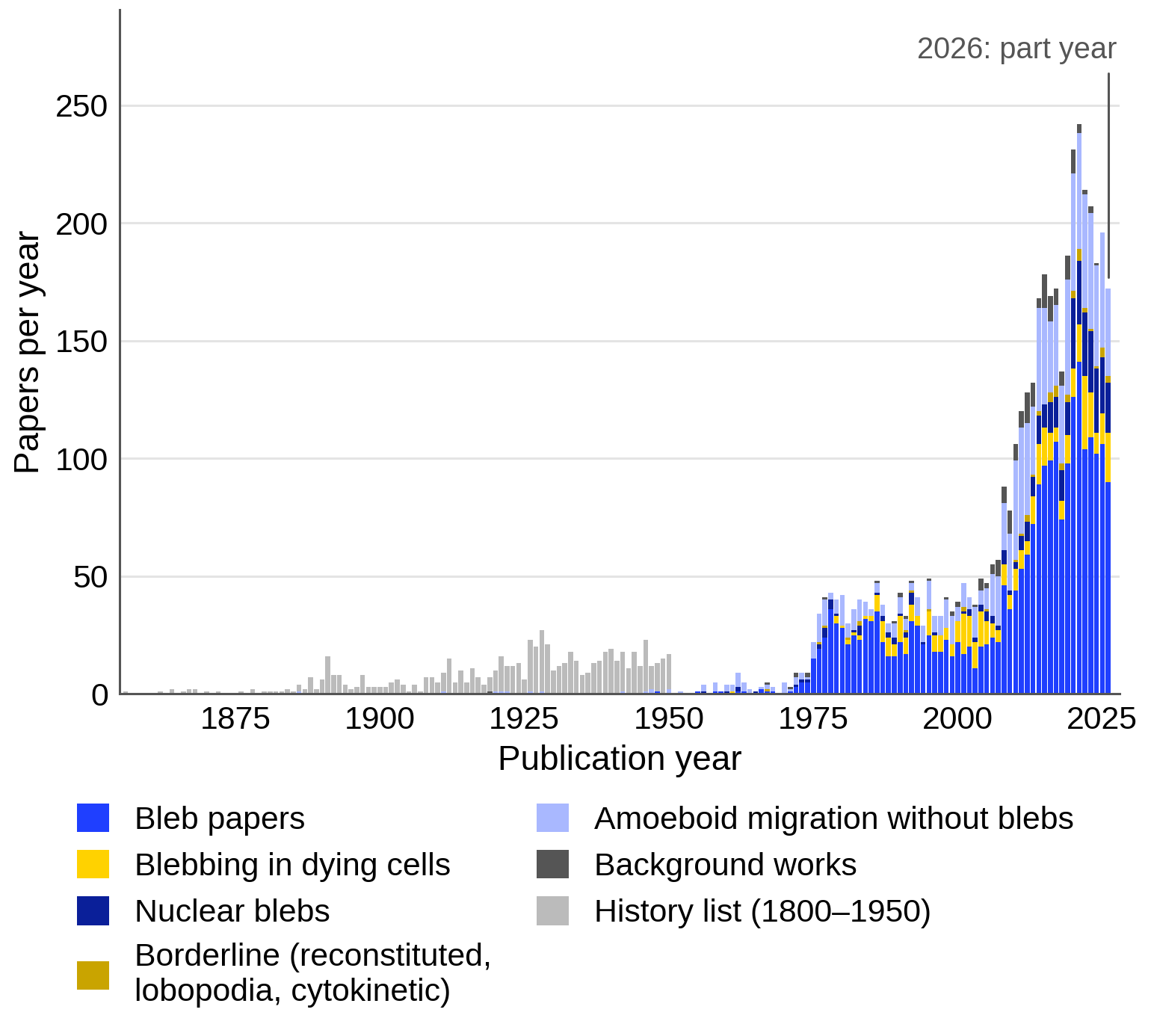
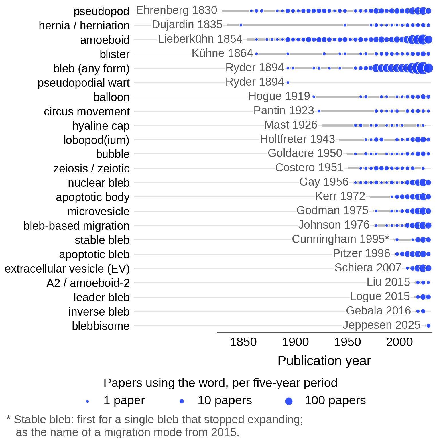
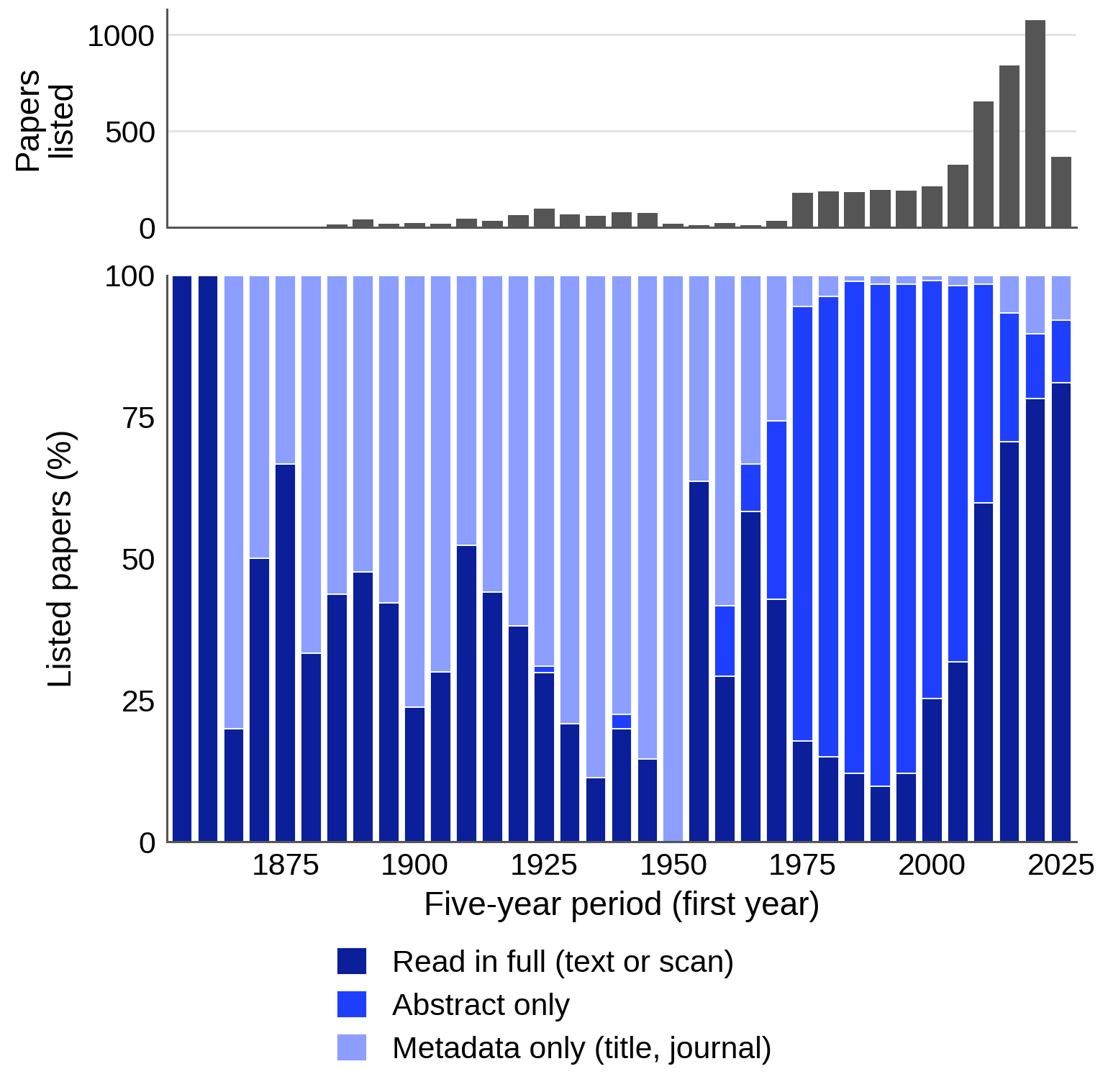
The bleb list holds 2,426 counted papers once two retracted papers are set aside. It grew from about 23 papers a year in 1975–2004 to 106 a year in 2015–2024, with a peak of 141 in 2021, so half of all bleb papers appeared in 2015 or later. The nuclear-bleb list, which concerns a different membrane, grew faster still, from 54 papers in 2005–2014 to 245 in 2015–2026.
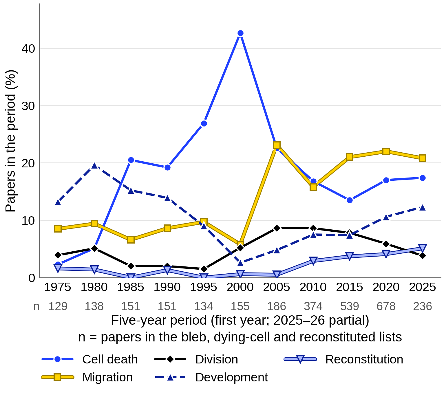
What the papers are about shifted twice. Cell death accounted for 10% of papers in 1975–1989, 30% in 1990–2004 and 43% in 2000–2004, the highest share of any setting in any period since 1975; since papers that use blebbing merely as a marker of death were excluded, these papers study the blebbing of dying cells. Migration then took over, rising from about 8% of papers before 2005 to 18% in 2005–2014 and 21% in 2015–2026, the most common of the five settings in every five-year period since 2015, while cell death fell back to 16%. Development fell from 16% in 1975–1989 to about 3% in 2000–2004 and has risen again, to 12% in 2025–2026, and reconstitution in cell-free or vesicle systems rose from 1% or less before 2005 to 4% in 2015–2026.
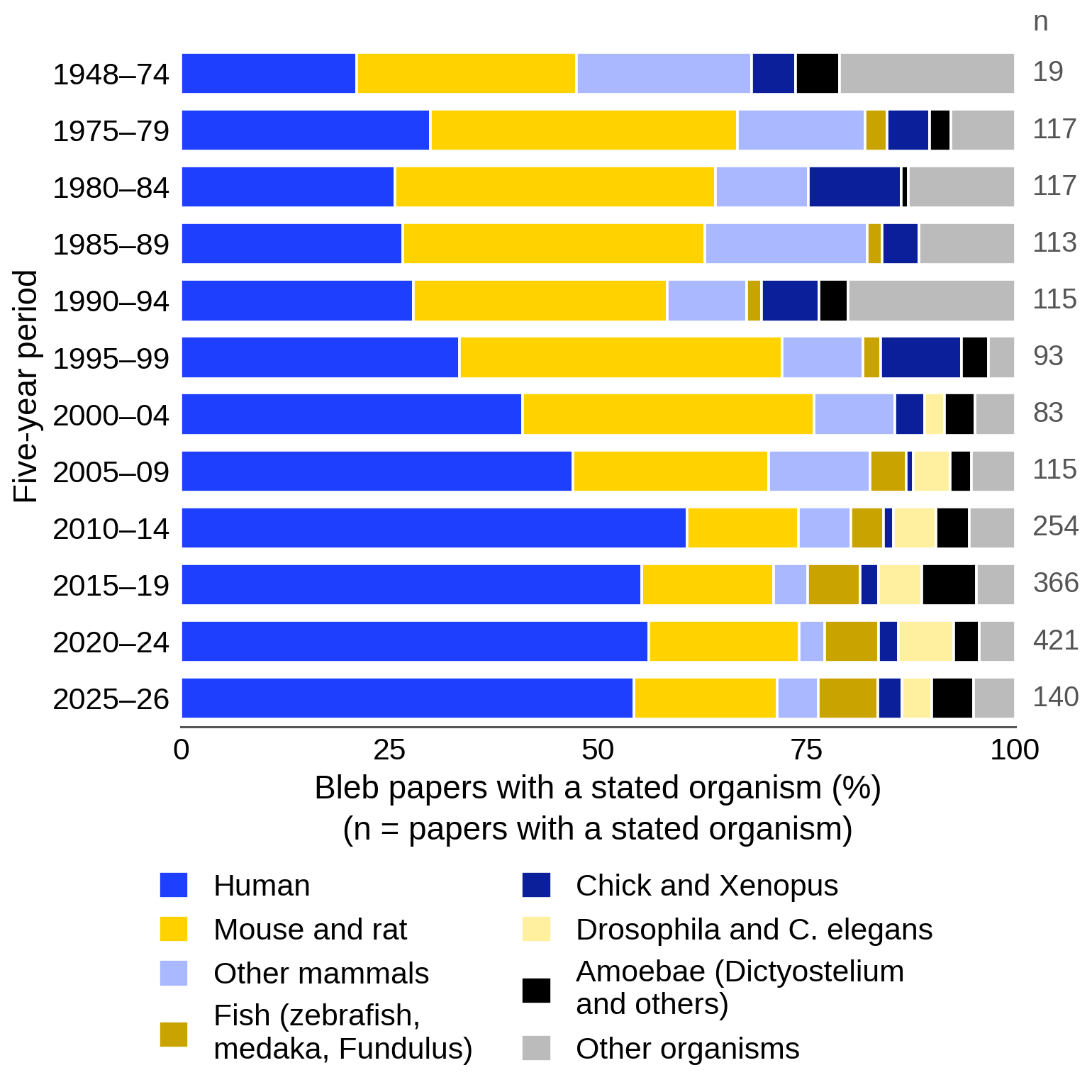
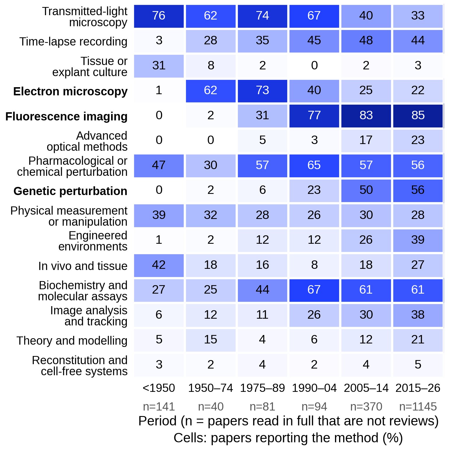
The systems and tools changed with the questions. Among bleb papers that name an organism, human cells rose from 27% in 1975–1989 to 55% in 2015–2026, mouse and rat cells fell from 37% to 17%, and fish, flies, worms and amoebae together grew from about 3% to about 17%. Among papers read in full, over the same periods, electron microscopy fell from 73% to 22%, while fluorescence imaging rose from 31% to 85%, genetic perturbation from 6% to more than half, engineered environments such as gels and microchannels from 12% to 39%, and modeling from 4% to 21%. The early method shares rest on only 81 papers read in full, so they are rough.
The vocabulary settled as the field grew. "Bleb" in some form occurs in 80% of the 86 full texts searched from 1975–1989 and in 91% of the 1,547 from 2015–2026, while "zeiosis" fell from 20% to 0.1%. Newer words name modes and products rather than shapes: phrases for bleb-based migration occur in 13% of the full texts from 2015–2026 and "extracellular vesicle" in 15%, and leader bleb, inverse bleb and blebbisome first appear in our notes between 2015 and 2025.
How far the counts can be trusted
The searches define the vocabulary. Because the lists were found with bleb words and four older terms, the dominance of "bleb" is partly built in, and papers that say only "blister", "bubble" or "lobopodium" enter mainly through full text or the citation check. Database coverage also changes: before about 1975 PubMed records rarely carry abstracts, so the jump of the bleb list from 14 papers in 1970–1974 to 124 in 1975–1979 is probably partly an artifact. The historical list was sorted from titles alone and is not comparable with the later lists.
Access is the largest source of bias. Searching inside articles reached only open-access papers, about a third of the bleb papers were found only that way, and the 5,303 papers without open full text were not examined at all. The rules that thinned the full-text hits may also have lost about one relevant paper in ten, a rough estimate from a test of ten papers. Only 15% of listed papers from 1975–2004 were read in full, against 71% of those from 2010–2024, and 70% of the records from before 1950 carry only a title and journal, so trends for the older periods rest mostly on abstracts. Growth in open access should not be mistaken for growth of the field.
Sorting has an expected error of about 5%, from the audit of 100 decisions, while the topic tags were only spot-checked. The tags follow the categories of one review (García-Arcos et al., 2024a ◆), so we checked them against how papers by other groups distribute. Between 55% and 74% of bleb papers in each period since 1975 carry only the code "function or phenotype": they report blebs, or what blebs do, without studying how blebs form. The counts are therefore a map of activity rather than of understanding.
What reviews up to 2018 predicted, and what happened
To judge how far reviews can guide expectations, we went back to 147 reviews, perspectives, books and theses dated 1764 to 2018, extracted 1,471 predictions and open questions, and scored each against later primary papers in the reading notes. Language-model readers did the scoring under written rules: only primary papers published after the source counted, and a later review could mark a point as still open but never as answered. About half of the items, 721, concerned matters the lists were not built to cover. Of the 750 that could be tested, 17% were realized, 43% partly realized, 38% not yet and 1.5% contradicted. For sources from 2005–2018, the first full answer came a median of 5 years after the review and the first partial answer a median of 4 years. "Not yet" means not found in these lists, and because the lists were partly assembled around the papers that answered the old questions, the record is flattered.
The clearest success concerned migration without adhesion. Charras and Paluch (2008) proposed that cells squeezed between surfaces could bleb forward without specific adhesion, whereas Lämmermann and Sixt (2009) argued that blebbing tumor cells draw their traction from residual adhesive protrusions. In 2015, confined cells of many types migrated fast on non-adhesive surfaces, driven by rearward cortical flow that grips the walls by friction (Bergert et al., 2015; Liu et al., 2015; Ruprecht et al., 2015). The switch is established for confined cells in vitro (M20), and 2015 alone brought the first answer to 47 of the old items, 32 of them through four papers on confined cells.
Other forecasts came true in part, often because a general question received a different answer in each system. Charras (2008) asked, for example, whether lipid flows into growing blebs. It did not in zebrafish germ cells, where invaginations of the surface unfolded instead (Goudarzi et al., 2017), but it flowed from base to tip in the stable leader blebs of melanoma cells (Jha et al., 2026). The answer depends on the kind of bleb, which the question had not separated.
Some questions remain open after nearly two decades. Reviews of 2006–2008 asked whether blebs in migrating cells start where the membrane detaches from an intact cortex, the actin layer beneath it, or where the cortex tears (rupture versus detachment). Reconstituted cortices in liposomes show that both routes are possible (Miyazaki et al., 2026), and in single examples from nucleus-free fragments of L929 cells the cortex tore before the bulge grew (Paluch et al., 2005). In Dictyostelium, however, one study saw the old cortex persist intact after each bleb (Zatulovskiy et al., 2014) and another found narrow gaps at inferred nucleation sites (Asante-Asamani et al., 2022). In our reading, a narrow gap is easy to miss in a survey, and the targeted analysis is correlative. Whether migrating vertebrate cells start blebs by rupture is an open question (M15).
Some kinds of forecast fared better than others. Calls for a new measurement, tool or experimental system were fully realized in 28% of cases, against 17% for other statements, probably because they usually describe work already under way. Statements about physical quantities such as tension, pressure or friction did better than those naming only molecules (26% against 18%), forecasts set in a living organism or tissue did worse (12% against 22% where no setting was named), and therapy, biomarker and clinical forecasts fared worst, with 4% realized and 65% not yet. Five of the 11 contradictions came from claims that one mechanism was universal or exclusive, such as the proposal that an adhesion–RhoA–ROCK–myosin II axis controls all migration in three dimensions (Petrie and Yamada, 2012), whose adhesion arm was contradicted by adhesion-free migration in confinement (Bergert et al., 2015). Forecasters also often answered their own questions: for 28 of 105 realized modern items, the first qualifying paper shared an author surname with the review.
Where current reviews expect the field to go
To see where the field is heading, we read the 323 reviews, perspectives, commentaries, books and theses from 2019–2026 in the lists, 31 of them only as abstracts and 8 from the record alone. Most mention blebs only in passing: about 110 are reviews of extracellular vesicles that repeat the textbook link between blebbing and vesicle release, most of the 26 on the nucleus use "bleb" for herniations of the nuclear envelope, about 50 are off-topic, and only 17 are centered on blebs. On bleb nucleation, membrane tension and theory, primary papers are ahead of the reviews. We grouped the directions that reviewers name into six themes and set each against the record of similar forecasts in the backward check, which describes kinds of forecasts, not the odds for any one.
Whether blebs are needed in living tissue. The direction named most often, by about fifteen reviews and commentaries, is to show that blebs are required in tissues, not merely present (Schick and Raz, 2022; Caruso and Logue, 2024; García-Arcos et al., 2024a ◆). The difficulty is that inhibiting myosin or ROCK, the usual way to stop blebs, also lowers the contractility this migration needs, so blebs could be a by-product (Schick and Raz, 2022). Perturbations aimed at the bleb itself are beginning to appear: chick germ cells carrying a dominant-negative form of the calcium channel Orai1 rarely blebbed and left the blood vessels about 40% less efficiently (4 embryos; Morita et al., 2026). Mouse primordial germ cells, in contrast, migrate with actin-rich protrusions and bleb only when actin polymerization is blocked (Goodwin et al., 2026). Forecasts set in a living organism were realized in only 12% of cases.
The physics of a single bleb. Roughly six to eleven reviews each ask how a bleb starts, where its extra membrane comes from, how water and pressure move as it grows, and whether the connectivity of the cortex controls it (Ikenouchi and Aoki, 2022; Fujii and Ikenouchi, 2024). Water flow and pressure are still inferred rather than measured: an osmotic model of bleb growth needs about twice the protein concentration difference that was measured (Fujii et al., 2026). Cortex connectivity as a control variable rests largely on one cell line (García-Arcos et al., 2024b ◆) and one liposome study. In the backward check, such mechanistic questions took longest to answer, a median of six to seven years, and the answers usually held in one system.
Rebuilding blebs from purified parts. A smaller group expects blebbing to be rebuilt from purified components, first as single blebs, then as blebs that retract and form again, and eventually as movement. Only one recent review discusses it (Lopes Dos Santos and Campillo, 2022). Vesicles with an actomyosin shell formed static blebs where the shell was less firmly anchored to the membrane (Loiseau et al., 2016), and laser cuts in a model cortex triggered blebs in at most 13% of liposomes (Sakamoto and Murrell, 2024), but none of these blebs has yet retracted by rebuilding its cortex (M15). Calls for a method, measurement or reconstituted system have the best record in the backward check, fully realized in 28–31% of cases.
Blebs as signaling sites, vesicles and fragments. About fifteen reviews and commentaries treat blebs as compartments that concentrate signals or leave the cell (Reichman-Fried and Raz, 2023; Di Vizio et al., 2023; García-Arcos et al., 2024a ◆), but the evidence is narrower than the attention. The view of blebs as signaling hubs rests mainly on septin-organized survival signaling in detached melanoma cells in vitro (Weems et al., 2023) and on polarized signaling modules in leader blebs (Jha et al., 2026). The expectation that blebs give rise to motile fragments in the body rests on stable blebs detaching in one confined cell line (García-Arcos et al., 2024b ◆) and on lung fragments whose bleb origin was not tested (Headley et al., 2016); here the forecast and the main evidence come from the same group. Blebbisomes, vesicles that keep blebbing for days, form in a retraction rather than from a bleb (Jeppesen et al., 2025). Of past forecasts of this kind, none of 43 testable items on signaling machinery and 6% of 193 on vesicles were fully realized.
Treating amoeboid cancer cells. Cancer reviews agree more closely than any other group. About ten expect amoeboid migration, the rounded, contractile and bleb-rich mode of some invading cancer cells, to become a drug target, because amoeboid cells gather at invasive fronts and depend on Rho–ROCK–myosin II contractility (Graziani et al., 2022; Jia et al., 2024; Ghose and Sanz-Moreno, 2026). ROCK inhibitors combined with drugs that block escape into other modes should then slow dissemination, although even Jia et al. (2024) judge clinical translation to be distant. Forecasts of this kind have the weakest record. Systemic ROCK inhibition reduced growth, invasion and lung metastasis in a mouse melanoma model (5 control and 4 treated mice; Rodriguez-Hernandez et al., 2020), but AT13148, a ROCK-targeting drug tested in a phase I cancer trial, was closed for adverse effects and lack of response (Graziani et al., 2022).
New ways to classify migration, and the origin of blebbing. About eight reviews expect migration to be classified by how force is generated and transmitted rather than by protrusion shape, because one cell can combine blebs with lamellipodia and the modes may form a continuum (Bodor et al., 2020; Alonso-Matilla et al., 2025; Duggan and Petrie, 2025). Choanoflagellates, the closest living relatives of animals, bleb when confined (Brunet et al., 2021), and one review proposes that migration without adhesions is older than migration driven by focal adhesions (Bodor et al., 2020). Past forecasts here held when they predicted variety, with new modes found within two to five years, and failed when they claimed generality. Whether blebs are older than lamellipodia, asked by Charras and Paluch (2008), is untested, so this expectation is an agenda rather than a forecast.
In our reading, the two checks together suggest how to read today's expectations. Partial answers tend to arrive within about four years, usually in one cell type or species. Calls for methods and measurements deserve more weight than forecasts of clinical benefit, and words such as "universal" or "the main determinant" deserve caution. The lists behind these counts can be downloaded from Paper lists.
References (38)
- Alonso-Matilla R, Provenzano PP, Odde DJ (2025) Physical principles and mechanisms of cell migration. npj Biol Phys Mech 2:2. doi:10.1038/s44341-024-00008-w
- Asante-Asamani E, Grange D, Rawal D et al. (2022) A role for myosin II clusters and membrane energy in cortex rupture for Dictyostelium discoideum. PLoS ONE e0265380. doi:10.1371/journal.pone.0265380
- Bergert M, Erzberger A, Desai RA et al. (2015) Force transmission during adhesion-independent migration. Nat Cell Biol 17:524–529. doi:10.1038/ncb3134
- Bodor DL, Pönisch W, Endres RG et al. (2020) Of cell shapes and motion: the physical basis of animal cell migration. Dev Cell 52:550–562. doi:10.1016/j.devcel.2020.02.013
- Brunet T, Albert M, Roman W et al. (2021) A flagellate-to-amoeboid switch in the closest living relatives of animals. eLife 10:e61037. doi:10.7554/elife.61037
- Caruso AP, Logue JS (2024) The biophysics of cell motility through mechanochemically challenging environments. Curr Opin Cell Biol 90:102404. doi:10.1016/j.ceb.2024.102404
- Charras GT (2008) A short history of blebbing. J Microsc 231:466–478. doi:10.1111/j.1365-2818.2008.02059.x
- Charras G, Paluch E (2008) Blebs lead the way: how to migrate without lamellipodia. Nat Rev Mol Cell Biol 9:730–736. doi:10.1038/nrm2453
- Di Vizio D, Schoppet M, Weeraratna A et al. (2023) Blebs and former blebs: from surface protrusions to extracellular vesicles in cancer signalling, anoikis resistance and beyond. J Extracell Biol 2:e112. doi:10.1002/jex2.112
- Duggan JJ, Petrie RJ (2025) The role of dynamic reciprocity in 3D cell migration: connecting cell and matrix mechanics to migratory plasticity. npj Biol Phys Mech 2:21. doi:10.1038/s44341-025-00027-1
- Fujii Y, Ikenouchi J (2024) Cytoplasmic zoning in membrane blebs. J Biochem 175:133–140. doi:10.1093/jb/mvad084
- Fujii Y, Sakai Y, Matsuzawa K et al. (2026) CaMKII nucleates an osmotic protein supercomplex to induce cellular bleb expansion. EMBO J 45:2433–2455. doi:10.1038/s44318-026-00703-5
- ◆ García-Arcos JM, Jha A, Waterman CM et al. (2024a) Blebology: principles of bleb-based migration. Trends Cell Biol 34:838–853. doi:10.1016/j.tcb.2024.02.009
- ◆ García-Arcos JM, Ziegler J, Grigolon S et al. (2024b) Rigidity percolation and active advection synergize in the actomyosin cortex to drive amoeboid cell motility. Dev Cell 59:2990–3007.e7. doi:10.1016/j.devcel.2024.06.023
- Ghose R, Sanz-Moreno V (2026) Amoeboid cancer cells at a glance. J Cell Sci 139:jcs264674. doi:10.1242/jcs.264674
- Goodwin K, Emrich TA, Arnold S et al. (2026) Primordial germ cells experience increasing physical confinement and DNA damage during migration in the mouse embryo. Sci Adv 12:eaec7919. doi:10.1126/sciadv.aec7919
- Goudarzi M, Tarbashevich K, Mildner K et al. (2017) Bleb expansion in migrating cells depends on supply of membrane from cell surface invaginations. Dev Cell 43:577–587.e5. doi:10.1016/j.devcel.2017.10.030
- Graziani V, Rodriguez-Hernandez I, Maiques O et al. (2022) The amoeboid state as part of the epithelial-to-mesenchymal transition programme. Trends Cell Biol 32:228–242. doi:10.1016/j.tcb.2021.10.004
- Headley MB, Bins A, Nip A et al. (2016) Visualization of immediate immune responses to pioneer metastatic cells in the lung. Nature 531:513–517. doi:10.1038/nature16985
- Ikenouchi J, Aoki K (2022) A Clockwork Bleb: cytoskeleton, calcium, and cytoplasmic fluidity. FEBS J 289:7907–7917. doi:10.1111/febs.16220
- Jeppesen DK, Sanchez ZC, Kelley NM et al. (2025) Blebbisomes are large, organelle-rich extracellular vesicles with cell-like properties. Nat Cell Biol 27:438–448. doi:10.1038/s41556-025-01621-0
- Jha A, Chandra A, Farahani PE et al. (2026) CD44 restricts EGFR mobility to polarize cytoskeletal signalling modules driving bleb-based migration. Nat Cell Biol 28:1408–1423. doi:10.1038/s41556-026-01981-1
- Jia W, Czabanka M, Broggini T (2024) Cell blebbing novel therapeutic possibilities to counter metastasis. Clin Exp Metastasis 41:817–828. doi:10.1007/s10585-024-10308-z
- Lämmermann T, Sixt M (2009) Mechanical modes of 'amoeboid' cell migration. Curr Opin Cell Biol 21:636–644. doi:10.1016/j.ceb.2009.05.003
- Liu YJ, Le Berre M, Lautenschlaeger F et al. (2015) Confinement and low adhesion induce fast amoeboid migration of slow mesenchymal cells. Cell 160:659–672. doi:10.1016/j.cell.2015.01.007
- Loiseau E, Schneider JAM, Keber FC et al. (2016) Shape remodeling and blebbing of active cytoskeletal vesicles. Sci Adv 2:e1500465. doi:10.1126/sciadv.1500465
- Lopes Dos Santos R, Campillo C (2022) Studying actin-induced cell shape changes using Giant Unilamellar Vesicles and reconstituted actin networks. Biochem Soc Trans 50:1527–1539. doi:10.1042/bst20220900
- Miyazaki M, Laboni FS, Kim T (2026) Reconstitution of actomyosin networks in cell-sized liposomes dissects distinct mechanisms of membrane blebbing and symmetry breaking. Sci Adv 12:eaed8818. doi:10.1126/sciadv.aed8818
- Morita M, Morimoto M, Ikenouchi J et al. (2026) Bleb-based extravasation uses conserved morphodynamics but divergent calcium control. Nat Commun 17:4422. doi:10.1038/s41467-026-71052-4
- Paluch E, Piel M, Prost J et al. (2005) Cortical actomyosin breakage triggers shape oscillations in cells and cell fragments. Biophys J 89:724–733. doi:10.1529/biophysj.105.060590
- Petrie RJ, Yamada KM (2012) At the leading edge of three-dimensional cell migration. J Cell Sci 125:5917–5926. doi:10.1242/jcs.093732
- Reichman-Fried M, Raz E (2023) Bleb protrusions help cancer cells to cheat death. Nature 615:402–403. doi:10.1038/d41586-023-00477-4
- Rodriguez-Hernandez I, Maiques O, Kohlhammer L et al. (2020) WNT11-FZD7-DAAM1 signalling supports tumour initiating abilities and melanoma amoeboid invasion. Nat Commun 11:5315. doi:10.1038/s41467-020-18951-2
- Ruprecht V, Wieser S, Callan-Jones A et al. (2015) Cortical contractility triggers a stochastic switch to fast amoeboid cell motility. Cell 160:673–685. doi:10.1016/j.cell.2015.01.008
- Sakamoto R, Murrell MP (2024) Active tension and membrane friction mediate cortical flows and blebbing in a model actomyosin cortex. Phys Rev Res 6:033024. doi:10.1103/PhysRevResearch.6.033024
- Schick J, Raz E (2022) Blebs—formation, regulation, positioning, and role in amoeboid cell migration. Front Cell Dev Biol 10:926394. doi:10.3389/fcell.2022.926394
- Weems AD, Welf ES, Driscoll MK et al. (2023) Blebs promote cell survival by assembling oncogenic signalling hubs. Nature 615:517–525. doi:10.1038/s41586-023-05758-6
- Zatulovskiy E, Tyson R, Bretschneider T et al. (2014) Bleb-driven chemotaxis of Dictyostelium cells. J Cell Biol 204:1027–1044. doi:10.1083/jcb.201306147