M111998–2003 · The regulated bleb, 1995–2003 · strongly supported
ROCK–myosin contraction drives blebbing in dying and in invading cells
Until the late 1990s, the blebbing of stressed and dying cells was widely seen as a non-specific response to injury. Studies in 1998–2003 showed instead that myosin II contraction drives it, that caspase cleavage of Rho-associated kinase I (ROCK I) switches this contraction on in apoptosis, and that the same Rho–ROCK pathway drives a rounded, blebbing mode of tumor-cell invasion. That ROCK-driven myosin contraction powers these blebs is strongly supported, although for invasion the evidence comes from cell lines in vitro.
Before
Hints of an active process existed. Burnham and Williams (1982) saw over-stimulated mouse pancreatic acini form blebs that cytochalasin B largely prevented, and suggested that “Elevated contractility may raise intracellular fluid pressure” (p. 211), without measuring either. Outside apoptosis, the ROCK inhibitor Y-27632 abolished the blebbing of 293T cells stimulated through the receptor EphA3 (Lawrenson et al., 2002). Still unknown were the motor that drives these blebs, how death signaling switches it on, and whether moving cells use the same machinery.
What was done
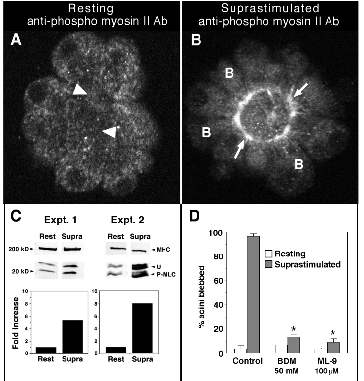
The first studies separated blebbing from death. In serum-deprived PC12 cells, z-VAD-FMK, an inhibitor of caspases, the proteases that execute programmed cell death, stopped death but not blebbing. Among kinase inhibitors, only those acting on myosin light-chain kinase (MLCK) reduced ongoing blebbing, and phosphorylation of the myosin regulatory light chain was 3–5-fold higher in blebbing cultures (Mills et al., 1998). Rat pancreatic acini, apparently not apoptotic, gave the same answer: they blebbed reversibly in supramaximal cholecystokinin, phosphorylated myosin ringed the bleb necks, and the myosin ATPase inhibitor BDM or the MLCK inhibitor ML-9 blocked blebbing (Torgerson and McNiven, 1998; figure).
How does apoptosis switch myosin on? Caspase cleavage made ROCK I 7.8 ± 0.5-fold more active (s.e.m., 5 experiments; Coleman et al., 2001), and in MCF-7 cells cleavage required caspase-3 (Sebbagh et al., 2001). In Jurkat cells triggered to die through Fas, Y-27632 abolished both light-chain phosphorylation and blebbing, whereas inactivating Rho affected neither (Sebbagh et al., 2001), and truncated ROCK I made cells bleb even with caspases blocked (Coleman et al., 2001; Sebbagh et al., 2001). Caspases thus activate ROCK I without Rho, and ROCK I activates myosin.
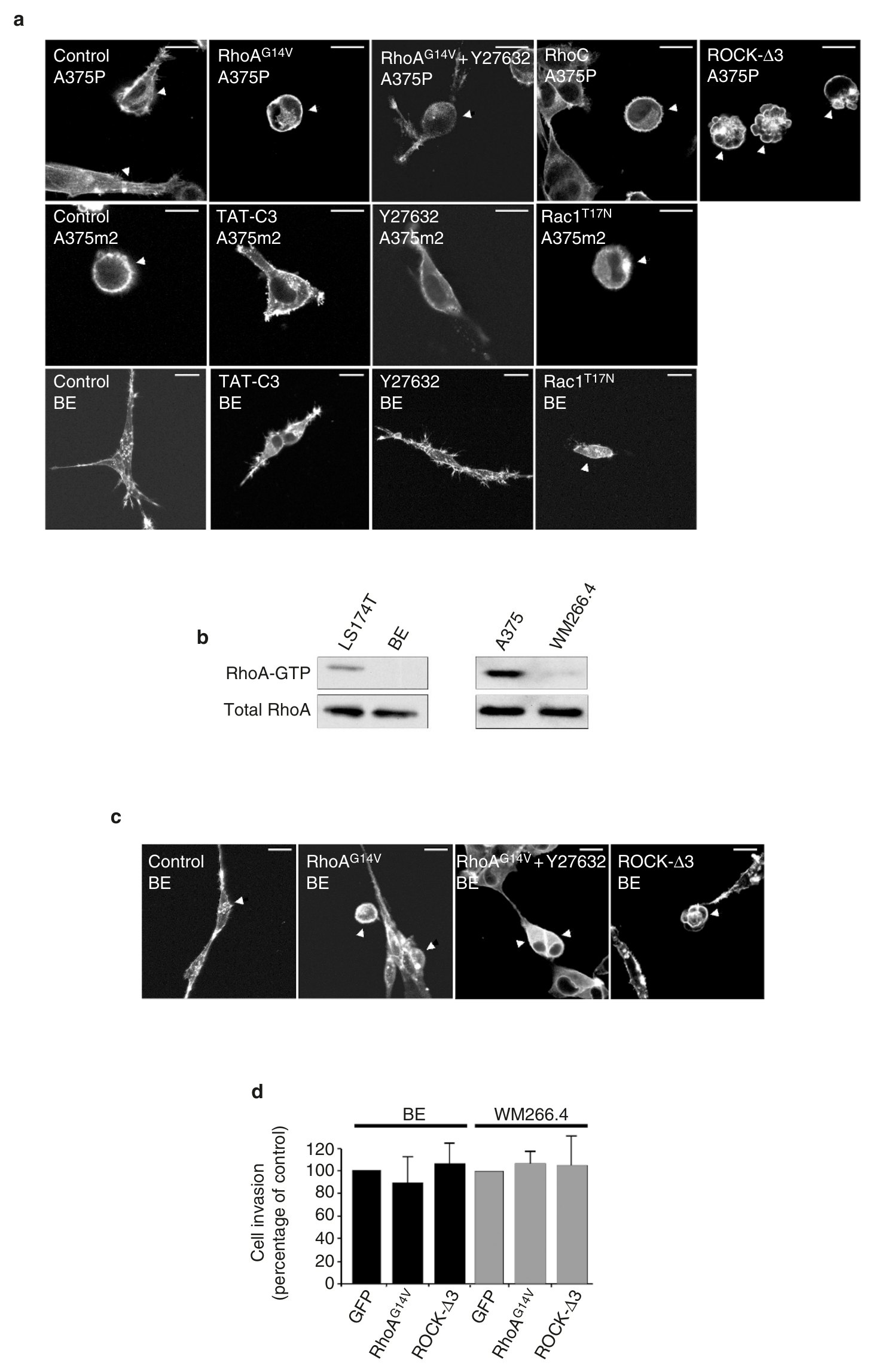
To ask whether moving cells use the same machinery, Sahai and Marshall (2003) compared five tumor lines invading Matrigel, a three-dimensional (3D) gel. Inhibiting Rho or ROCK almost abolished invasion by two rounded, blebbing lines but spared elongated SW962 cells (table), and active ROCK I made A375P cells round and blebbing (figure). Protease inhibitors did not reduce invasion by the rounded lines but made elongated BE cells invade with a rounded shape, which Y-27632 then cut sharply. The authors proposed that contractility raises hydrostatic pressure to push out blebs, but did not measure pressure or bleb dynamics.
| Quantity | Value | System | Source |
|---|---|---|---|
| Inhibition of ongoing blebbing at 1 h | staurosporine 89%, KT5926 39% (text); ML-9 94–99% at 80 µM (mean ± s.e.m., 3 experiments) | PC12, caspase-inhibited | Mills et al., 1998, Fig. 5 |
| Invasion with Y-27632 (10 µM), depth ≥12 µm | A375m2 3.8; LS174T 1.0; WM266.4 34; BE 78; SW962 157 (% of control; mean of 3 experiments, ≥1,000 cells each; no error given) | Transwell Matrigel | Sahai and Marshall, 2003, Table 1 |
| Invasion, depth ≥8 µm: Y-27632 / protease inhibitors / both | A375m2 12 / 150 / 6; LS174T 13 / 250 / 9; BE 81 / 115 / 16 (% of control; 3 experiments) | Transwell Matrigel | Sahai and Marshall, 2003, Table 3 |
| Later: apoptotic cells with surface blebs, control vs ROCK1−/− clones C1, C2, C3 | ≈100 vs ≈4, ≈89, ≈84% (read from plot; s.e.m., 3 experiments) | Jurkat, anti-Fas | Tixeira et al., 2020, Fig. 2e |
What it changed
The work turned blebbing into the output of a defined pathway shared by dying and moving cells. Mills et al. (1999) named myosin II, contracting against focally weakened membrane–cortex links, as the force in apoptotic and non-apoptotic blebbing alike, and Fackler and Grosse (2008) treated Rho–ROCK–myosin as the core machinery of both. Genetic evidence was partial: of three ROCK1-knockout Jurkat clones, one almost lost apoptotic blebbing and two lost part of it (Tixeira et al., 2020; table).
Tumor cells also proved able to switch between rounded and elongated movement. In A375M2 cells, ROCK inhibition made rounded cells elongated without slowing them, and depletion of the GTPase Rac left only amoeboid movement (Sanz-Moreno et al., 2008). This plasticity, the mesenchymal–amoeboid transition, became a standard frame for tumor invasion (Lämmermann and Sixt, 2009).
Later work and open questions
In our reading, several apparently contradictory findings reflect differences between cell types and stimuli. Blebbing continued without caspase activity in PC12 cells, where it fell when Rho was inhibited (Mills et al., 1998), and in cisplatin-treated Rat-1 cells, where it needed the stress kinase p38 (Deschesnes et al., 2001). In NIH 3T3 fibroblasts and Jurkat cells, by contrast, it depended on caspases and not on Rho (Coleman et al., 2001; Sebbagh et al., 2001); Sebbagh et al. suggested that adherent cells use Rho as they round up. In endothelial cells treated with combretastatin A-4-phosphate, inhibitors of MLCK and of myosin even increased blebbing (Kanthou and Tozer, 2002).
Invasion, too, depends on the environment. In cross-linked native collagen, tumor cells needed the membrane protease MT1-MMP to invade (Sabeh et al., 2009), so protease-free invasion in Matrigel cannot be assumed in tissue. In rat mammary tumors in mice, ROCK inhibition cut the number of moving tumor cells by about 70% and a broad metalloprotease inhibitor by only about a fifth (no statistics given), but in collagen gels these cells led with an actin-rich front, and blebs are not mentioned (Wyckoff et al., 2006). On flat collagen, unlike on plastic, Y-27632 slowed A375M2 cells (Tozluoğlu et al., 2013).
Two questions remain open. What ends apoptotic blebbing, given that truncated ROCK I sustains blebbing for hours whereas apoptotic blebbing of NIH 3T3 cells lasts about 20–30 min (Coleman et al., 2001)? And do blebs themselves give rounded tumor cells their invasive advantage, or does ROCK-driven contractility act otherwise? No perturbation yet removes blebs selectively (Guzman et al., 2020). See also Dying and injured cells and Migration.
References (18)
- Burnham DB, Williams JA (1982) Effects of high concentrations of secretagogues on the morphology and secretory activity of the pancreas: a role for microfilaments. Cell Tissue Res 222:201–212. doi:10.1007/bf00218300
- Coleman ML, Sahai EA, Yeo M et al. (2001) Membrane blebbing during apoptosis results from caspase-mediated activation of ROCK I. Nat Cell Biol 3:339–345. doi:10.1038/35070009
- Deschesnes RG, Huot J, Valerie K et al. (2001) Involvement of p38 in apoptosis-associated membrane blebbing and nuclear condensation. Mol Biol Cell 12:1569–1582. doi:10.1091/mbc.12.6.1569
- Fackler OT, Grosse R (2008) Cell motility through plasma membrane blebbing. J Cell Biol 181:879–884. doi:10.1083/jcb.200802081
- Guzman A, Avard RC, Devanny AJ et al. (2020) Delineating the role of membrane blebs in a hybrid mode of cancer cell invasion in three-dimensional environments. J Cell Sci 133:jcs236778. doi:10.1242/jcs.236778
- Kanthou C, Tozer GM (2002) The tumor vascular targeting agent combretastatin A-4-phosphate induces reorganization of the actin cytoskeleton and early membrane blebbing in human endothelial cells. Blood 99:2060–2069. doi:10.1182/blood.v99.6.2060
- Lämmermann T, Sixt M (2009) Mechanical modes of ‘amoeboid’ cell migration. Curr Opin Cell Biol 21:636–644. doi:10.1016/j.ceb.2009.05.003
- Lawrenson ID, Wimmer-Kleikamp SH, Lock P et al. (2002) Ephrin-A5 induces rounding, blebbing and de-adhesion of EphA3-expressing 293T and melanoma cells by CrkII and Rho-mediated signalling. J Cell Sci 115:1059–1072. doi:10.1242/jcs.115.5.1059
- Mills JC, Stone NL, Erhardt J et al. (1998) Apoptotic membrane blebbing is regulated by myosin light chain phosphorylation. J Cell Biol 140:627–636. doi:10.1083/jcb.140.3.627
- Mills JC, Stone NL, Pittman RN (1999) Extranuclear apoptosis: the role of the cytoplasm in the execution phase. J Cell Biol 146:703–708. doi:10.1083/jcb.146.4.703
- Sabeh F, Shimizu-Hirota R, Weiss SJ (2009) Protease-dependent versus -independent cancer cell invasion programs: three-dimensional amoeboid movement revisited. J Cell Biol 185:11–19. doi:10.1083/jcb.200807195
- Sahai E, Marshall CJ (2003) Differing modes of tumour cell invasion have distinct requirements for Rho/ROCK signalling and extracellular proteolysis. Nat Cell Biol 5:711–719. doi:10.1038/ncb1019
- Sanz-Moreno V, Gadea G, Ahn J et al. (2008) Rac activation and inactivation control plasticity of tumor cell movement. Cell 135:510–523. doi:10.1016/j.cell.2008.09.043
- Sebbagh M, Renvoizé C, Hamelin J et al. (2001) Caspase-3-mediated cleavage of ROCK I induces MLC phosphorylation and apoptotic membrane blebbing. Nat Cell Biol 3:346–352. doi:10.1038/35070019
- Tixeira R, Phan TK, Caruso S et al. (2020) ROCK1 but not LIMK1 or PAK2 is a key regulator of apoptotic membrane blebbing and cell disassembly. Cell Death Differ 27:102–116. doi:10.1038/s41418-019-0342-5 (online 2019)
- Torgerson RR, McNiven MA (1998) The actin-myosin cytoskeleton mediates reversible agonist-induced membrane blebbing. J Cell Sci 111:2911–2922. doi:10.1242/jcs.111.19.2911
- Tozluoğlu M, Tournier AL, Jenkins RP et al. (2013) Matrix geometry determines optimal cancer cell migration strategy and modulates response to interventions. Nat Cell Biol 15:751–762. doi:10.1038/ncb2775
- Wyckoff JB, Pinner SE, Gschmeissner S et al. (2006) ROCK- and myosin-dependent matrix deformation enables protease-independent tumor-cell invasion in vivo. Curr Biol 16:1515–1523. doi:10.1016/j.cub.2006.05.065