M171883–2025 · When blebs leave the cell · strongly supported
Blebbing as a route to cell fragmentation
Cells can lose pieces of themselves, and some pieces begin as blebs that detach, usually without a nucleus, and then move or carry cargo to other cells; other fragments keep blebbing once free. Bleb detachment is strongly supported as one route to fragments: it has been imaged in several systems, although the neck that parts has rarely been seen. How often blebs detach in tissue is not known, and many fragments form in other ways.
Before
That a piece of cytoplasm can move without a nucleus is a seminal observation of the nineteenth century. Cutting amoebae into pieces, a method called merotomy, Hofer (1889) found that anucleate pieces moved normally for 15–20 min and then weakly but without stopping, and lived 9–10 days on average.
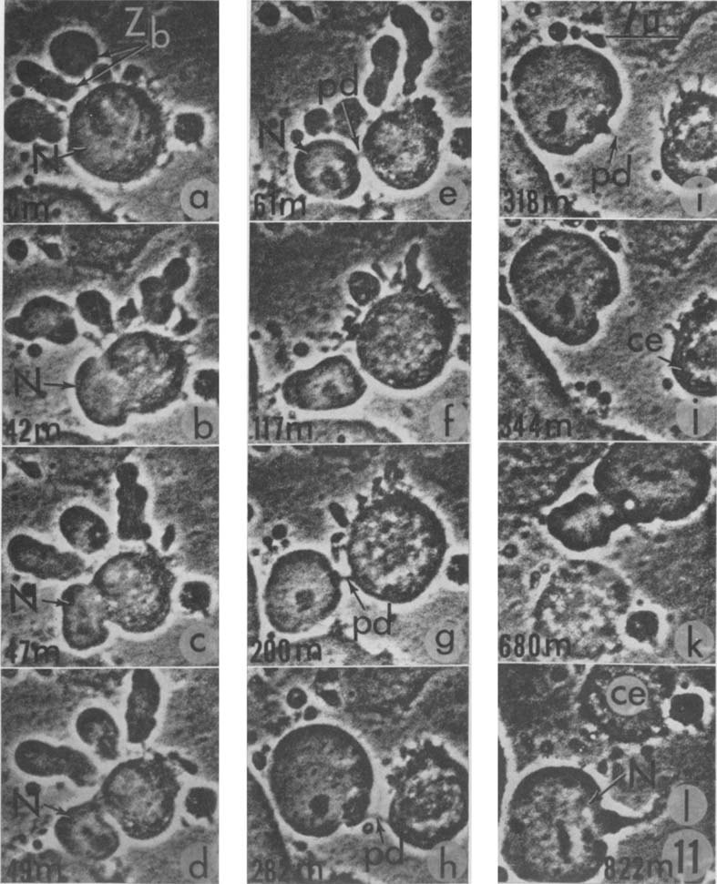
Detachment itself was watched in living cells. Holtfreter (1946) saw clear, granule-free processes of alkali-damaged amphibian embryonic cells pinch off as amoeboid vesicles, and anucleate fragments crawl for up to 7 days, although he did not call these processes blebs. KB carcinoma cells flattened under cellophane blebbed so actively that they seemed to boil, and some of their zeiotic blebs broke up into droplets of cytoplasm (Rose et al., 1963). Later films showed the stalk of such a bleb parting in three events, and one HeLa bleb carrying the nucleus kept moving for 11 h (Rose, 1966). Anecdotal as they are, these events show a bleb detaching and the freed piece moving.
Not every fragment moves, however: fibroblast microplasts made with cytochalasin blebbed or ruffled for at least 8 h but never translocated, so Albrecht-Buehler (1980) inferred that locomotion needs an additional supervising mechanism.
What was done
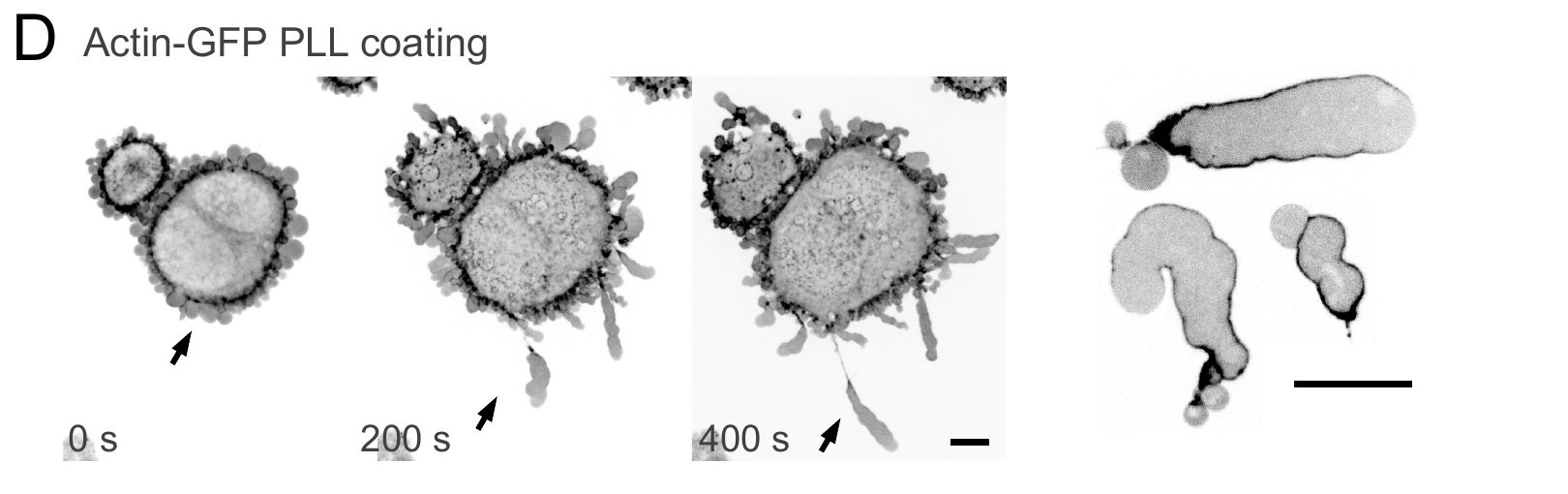
The clearest images of a bleb becoming a motile fragment come from confined HeLa cells. On poly-L-lysine, their stable blebs, large persistent blebs that can lead the cell, pulled away on a thinning neck until it ruptured, and the freed fragments moved for hours at 5–15 µm/min (García-Arcos, 2020 ◆; García-Arcos et al., 2024b ◆). These anucleate fragments, called cytokineplasts, kept the actin-poor front of stable blebs and squeezed through collagen (García-Arcos et al., 2024b ◆). Liu et al. (2015) had mentioned, without data, that shed large blebs moved like stable-bleb cells. The evidence comes from one laboratory and one cell line.
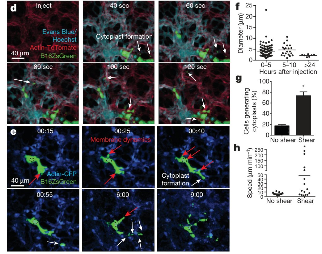
A second system showed fragmentation in a living animal. Melanoma cells injected into mice lodged in lung capillaries and by 40 s “began to shed microscale blebs” (Headley et al., 2016, p. 513). Shedding fell about fourfold without shear and did not need caspases, and anucleate cytoplasts soon made up most of the tumor material in the lung. With only cytoplasmic and nuclear labels, the neck and the bleb identity of these fragments could not be resolved.
Conversely, some fragments bleb without having come from a bleb. Blebbisomes, anucleate vesicles averaging 9–15 µm across, bleb for at least 72–96 h, yet in the one study of their origin they formed during a retraction of the cell and separated when a membrane nanotube was severed (Jeppesen et al., 2025); this route is proposed.
| Quantity | Value | System | Source |
|---|---|---|---|
| Fragment diameter | mean 5 µm; 0.5 to >25 µm stated (plot maximum ≈23 µm) | B16 melanoma cells, mouse lung | Headley et al., 2016, Fig. 1f |
| Cytokineplast speed | 5–15 µm/min, for hours | HeLa, 3 µm confinement | García-Arcos, 2020 ◆ |
| Blebbisome diameter | 8.92 ± 0.8 to 14.9 ± 3.0 µm (s.e.m., n = 167–236) | three cell lines | Jeppesen et al., 2025, Fig. 1b |
| Apoptotic cells fragmenting, pannexin 1 blocked vs not | ≈96% vs ≈6% (355, 454 cells; n = 3) | Jurkat cells | Poon et al., 2014, Fig. 2e |
What it changed
Fragments turned out to carry signals and cargo between cells. In the lung, myeloid cells took up the tumor cytoplasts, dendritic cells carried tumor material to the lymph node, and depleting conventional dendritic cells raised metastases sixfold (Headley et al., 2016).
Motile fragments also answered Albrecht-Buehler’s inference. Heat-made fragments of leukocytes migrated, as reported without data (Keller and Bessis, 1975), keratocyte fragments crawled persistently without a nucleus, centrioles or most microtubules (Euteneuer and Schliwa, 1984), and so did bleb-derived cytokineplasts (García-Arcos et al., 2024b ◆). That fragments without a nucleus can bleb and move is therefore established, although why his microplasts stayed in place was not tested. More is on the Fragments page.
Later work and open questions
Later papers, including two of ours, describe the lung fragments as bleb-derived. One cites a study “in human xenograft tumors” in which cytoplasts “were formed from large blebs” (García-Arcos et al., 2024b ◆, p. 3004), and the other has fragments “originating from stable blebs” (García-Arcos et al., 2024a ◆, p. 850). Headley et al. (2016) did call the fragments blebs, but they filmed injected mouse B16 cells; human cells appear only in a flow count, and neither xenografts nor stable blebs are mentioned. Whether the lung fragments are blebs was not tested.
Dying cells show that blebbing and fragmentation can be separated. With pannexin 1 channels blocked, blebs at the tips of apoptopodia, thin protrusions of dying cells, became apoptotic bodies (Poon et al., 2014). Yet bodies also formed from beaded apoptopodia when blebbing was blocked (Atkin-Smith et al., 2015), and phagocytes in zebrafish split apoptotic cells even with Rho-associated kinase (ROCK) inhibited (Santavanond et al., 2025).
Several questions remain open. No study has imaged the severing of a bleb neck with membrane and cortex markers; in our map of 195 papers on fragmentation, 19 image a bleb detaching and 101 only propose it. How often stable blebs detach in three-dimensional matrix and in tissue is an open question, and no criteria yet separate bleb-derived vesicles from large oncosomes and blebbisomes.
References (16)
- Albrecht-Buehler G (1980) Autonomous movements of cytoplasmic fragments. Proc Natl Acad Sci USA 77:6639–6643. doi:10.1073/pnas.77.11.6639
- Atkin-Smith GK, Tixeira R, Paone S et al. (2015) A novel mechanism of generating extracellular vesicles during apoptosis via a beads-on-a-string membrane structure. Nat Commun 6:7439. doi:10.1038/ncomms8439
- Euteneuer U, Schliwa M (1984) Persistent, directional motility of cells and cytoplasmic fragments in the absence of microtubules. Nature 310:58–61. doi:10.1038/310058a0
- ◆ García-Arcos JM (2020) Stabilization and motility mechanism of blebs in cancer cells. PhD thesis, Université de Paris (Institut Curie). doi:10.70675/a35575c7z85b5z4ad1z98acz50f8d65ebbac
- ◆ García-Arcos JM, Jha A, Waterman CM et al. (2024a) Blebology: principles of bleb-based migration. Trends Cell Biol 34:838–853. doi:10.1016/j.tcb.2024.02.009
- ◆ García-Arcos JM, Ziegler J, Grigolon S et al. (2024b) Rigidity percolation and active advection synergize in the actomyosin cortex to drive amoeboid cell motility. Dev Cell 59:2990–3007.e7. doi:10.1016/j.devcel.2024.06.023
- Headley MB, Bins A, Nip A et al. (2016) Visualization of immediate immune responses to pioneer metastatic cells in the lung. Nature 531:513–517. doi:10.1038/nature16985
- Hofer B (1889) Experimentelle Untersuchungen über den Einfluss des Kerns auf das Protoplasma. Jena Z Naturwiss 24:105–176.
- Holtfreter J (1946) Structure, motility and locomotion in isolated embryonic amphibian cells. J Morphol 79:27–62. doi:10.1002/jmor.1050790103
- Jeppesen DK, Sanchez ZC, Kelley NM et al. (2025) Blebbisomes are large, organelle-rich extracellular vesicles with cell-like properties. Nat Cell Biol 27:438–448. doi:10.1038/s41556-025-01621-0
- Keller HU, Bessis M (1975) Migration and chemotaxis of anucleate cytoplasmic leukocyte fragments. Nature 258:723–724. doi:10.1038/258723a0
- Liu YJ, Le Berre M, Lautenschlaeger F et al. (2015) Confinement and low adhesion induce fast amoeboid migration of slow mesenchymal cells. Cell 160:659–672. doi:10.1016/j.cell.2015.01.007
- Poon IKH, Chiu YH, Armstrong AJ et al. (2014) Unexpected link between an antibiotic, pannexin channels and apoptosis. Nature 507:329–334. doi:10.1038/nature13147
- Rose GG (1966) Zeiosis. I. Ejection of cell nuclei into zeiotic blebs. J R Microsc Soc 86:87–102. doi:10.1111/j.1365-2818.1966.tb05328.x
- Rose GG, Cattoni M, Pomerat CM (1963) Observations on oral tumors in vitro. 1. Zeiosis versus the nucleus and endoplasmic reticulum in strain KB. J Dent Res 42:38–53. doi:10.1177/00220345630420014401
- Santavanond JP, Atkin-Smith GK, Lozano-González I et al. (2025) Resident phagocytes promote non-cell-autonomous fragmentation of apoptotic cells. Sci Adv 11:eadz5264. doi:10.1126/sciadv.adz5264