M081973–1975 · Blebs on cultured animal cells, 1958–1984 · strongly supported
Blebbing on living, dividing cells follows cortical contraction
In the early 1970s blebs were familiar from injured, poisoned and dividing cells and were usually explained by a weakened or relaxed cell surface. Work in 1973–1975 showed that healthy cultured cells carry blebs, mostly in G1, the growth phase after division, and that blebbing induced by cytochalasin D, or zeiosis, needs an energy-consuming contraction, proposed to open gaps in the cortex, the actin-rich layer under the membrane. The contraction and its energy dependence are strongly supported, though by one group using several cell lines and inhibitors; the gaps-plus-pressure mechanism and the cell-cycle pattern remain proposed.
Before
Costero and Pomerat (1951, p. 432) had coined zeiosis (“to boil over”) for blebs on cultured neurons, but blebbing still had no agreed cause. Three explanations competed: a fall in ATP relaxing the surface, a loss of divalent cations weakening patches that pressure then blows out, and material released from the nucleus (Dornfeld and Owczarzak, 1958; Price, 1967; M07). None gave the cell an active role, and cytochalasins were thought to act by relaxing cells (as stated by Miranda et al., 1974a).
What was done
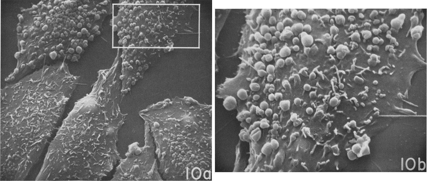
The first step was descriptive: Porter et al. (1973) synchronized Chinese hamster ovary (CHO) cells and imaged them through the cycle by scanning electron microscopy (SEM). By mid-G1 the cells carried many small spherical blebs among their microvilli; most S-phase cells lacked them, and by late S and G2 they had essentially gone (figure). Blebs thus belonged to healthy cycling cells, though they went uncounted.
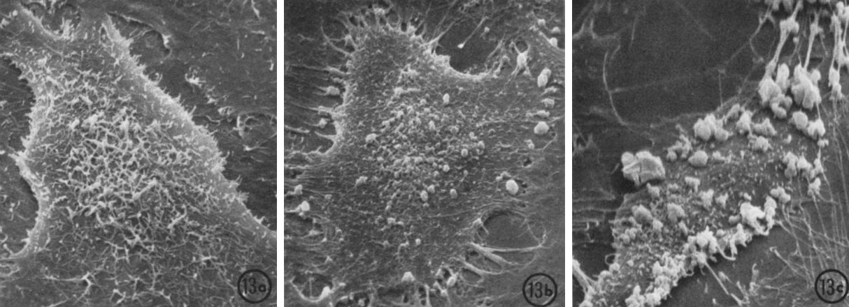
The second step was experimental: Miranda et al. (1974a) exposed six cell lines to cytochalasin D and scored 1,000 fixed cells per point for zeiosis. Within 2–3 min, cells contracted and formed the first knobs of endoplasm, the inner, organelle-rich cytoplasm (figure). HeLa and HEp2 cells needed doses about six- to eightfold lower than Vero and L cells, and G1 HeLa cells responded most, echoing the pattern seen without the drug (table). The decisive result came from metabolic poisons: 2-deoxyglucose and dinitrophenol, which deplete ATP, blocked contraction. Contraction, the authors concluded, was the drug’s primary, active effect, necessary but not sufficient for knob formation, and zeiosis was “only partly the result of increased intracellular pressure” (p. 494).
Sections suggested how contraction could produce knobs. Within 4 min of cytochalasin D, the filament mesh under the free surface gathered into compact pads with endoplasm bulging between them, and metabolic poisons prevented both. Because heavy meromyosin, a myosin fragment that decorates actin, still bound the pads, the drug was read as compacting cortical actin rather than destroying it (Miranda et al., 1974b). In nine lines, inhibitors that blocked contraction also prevented zeiosis, as did dibutyryl cyclic AMP, cooling and hypertonic sorbitol, and compacted filaments lay under the knobs and in their stalks but not in the bulbs (Godman et al., 1975; table). The authors summarized the process as “herniation of endoplasm through undefended places of the cortex” (p. 644).
| Quantity | Value (units, error, n) | System | Source |
|---|---|---|---|
| Bleb diameter | 0.2–1.5 µm (range; n not stated) | CHO, G1, SEM | Porter et al., 1973, Fig. 10a caption |
| Onset of contraction and first knobs | 2–3 min | several lines, 0.25–0.50 µg/ml cytochalasin D | Miranda et al., 1974a, Results; Fig. 13b |
| Dose for 50% zeiosis, 1 h | ≈0.05–0.065 µg/ml HeLa, HEp2; ≈0.4 Vero, L; not reached, MDBK (read from plot; 1,000 cells/point); tabulated 0.04–0.05 vs 0.25–0.35 | monolayers | Miranda et al., 1974a, Fig. 3; Godman et al., 1975, Table IV |
| Cells with zeiosis by phase | ≈61–70% G1 vs ≈22–30% S/G2 (read from plot; corrected for controls; n not stated) | synchronized HeLa, 0.2 µg/ml cytochalasin D, 10 min | Miranda et al., 1974a, Fig. 6 |
| Cells with zeiosis ± inhibitor | none 70%; 2-deoxyglucose 2%; dinitrophenol 2%; antimycin A 4%; CCCP 3%; KCN 39%; iodoacetate 48%; azide 54% (1,000 cells; single values) | HeLa, 0.25 µg/ml cytochalasin D, 15 min | Godman et al., 1975, Table II |
What it changed
The cytochalasin papers recast drug-induced blebbing as an energy-consuming result of cortical contraction rather than of injury or relaxation, which in our reading inverted the older ATP-depletion explanation. They also proposed a sequence that later work tested piece by piece: contraction raises intracellular pressure, the cortex opens and endoplasm herniates (Miranda et al., 1974b; Godman et al., 1975). A qualitative study later saw actin beneath clusters of knobs in another cell type treated with another cytochalasin (Watanabe et al., 1986).
Each element returned two decades later with better tools. Membrane was seen separating from a cortex left behind, which gave the “gap” a structural meaning (Keller and Eggli, 1998; M10). Inhibitors of myosin ATPase, myosin light-chain kinase or Rho-associated kinase (ROCK) blocked blebbing, placing myosin II at the center of the contraction (Torgerson and McNiven, 1998; Verhoef et al., 2003; M11), and inhibiting contraction over one part of a cell suppressed blebbing on that side only (Charras et al., 2005).
Later work and open questions
The cell-cycle pattern depended on culture conditions. Sparse CHO cells stayed blebbed into S and G2 and lost their blebs as they flattened and touched one another, suggesting that contact rather than cycle phase removed them (Rubin and Everhart, 1973), yet blebbed and smooth CHO cells were also found with or without contact (Porter et al., 1974). CHO is also a transformed line, and Porter et al. (1973) linked prolonged blebbing to transformation.
The mechanism rests on limited measurements. Zeiosis was scored without a stated criterion, error bars or replicates, contraction was inferred from cell area, and pressure was never measured. Because the inhibitors block contraction itself, they cannot tell whether knobs form because contraction opens gaps or because it raises pressure, and hypertonic sorbitol, the only indirect test of pressure, was reported without counts. Nor is it clear how typical the drug-induced knobs are, since they persisted for days on filament-filled stalks, unlike most zeiotic blebs (Godman et al., 1975).
Three questions remain open: why G1 cells and epithelial lines herniate more readily, how much of zeiosis comes from raised pressure rather than from gaps in the cortex (M12), and how a drug that binds actin can cause a sustained, energy-dependent contraction.
References (14)
- Charras GT, Yarrow JC, Horton MA et al. (2005) Non-equilibration of hydrostatic pressure in blebbing cells. Nature 435:365–369. doi:10.1038/nature03550
- Costero I, Pomerat CM (1951) Cultivation of neurons from the adult human cerebral and cerebellar cortex. Am J Anat 89:405–467. doi:10.1002/aja.1000890304
- Dornfeld EJ, Owczarzak A (1958) Surface responses in cultured fibroblasts elicited by ethylenediaminetetraacetic acid. J Biophys Biochem Cytol 4:243–250. doi:10.1083/jcb.4.3.243
- Godman GC, Miranda AF, Deitch AD et al. (1975) Action of cytochalasin D on cells of established lines. III. Zeiosis and movements at the cell surface. J Cell Biol 64:644–667. doi:10.1083/jcb.64.3.644
- Keller H, Eggli P (1998) Protrusive activity, cytoplasmic compartmentalization, and restriction rings in locomoting blebbing Walker carcinosarcoma cells are related to detachment of cortical actin from the plasma membrane. Cell Motil Cytoskeleton 41:181–193. doi:10.1002/(SICI)1097-0169(1998)41:2<181::AID-CM8>3.0.CO;2-H
- Miranda AF, Godman GC, Deitch AD et al. (1974a) Action of cytochalasin D on cells of established lines. I. Early events. J Cell Biol 61:481–500. doi:10.1083/jcb.61.2.481
- Miranda AF, Godman GC, Tanenbaum SW (1974b) Action of cytochalasin D on cells of established lines. II. Cortex and microfilaments. J Cell Biol 62:406–423. doi:10.1083/jcb.62.2.406
- Porter K, Prescott D, Frye J (1973) Changes in surface morphology of Chinese hamster ovary cells during the cell cycle. J Cell Biol 57:815–836. doi:10.1083/jcb.57.3.815
- Porter KR, Puck TT, Hsie AW et al. (1974) An electron microscope study of the effects of dibutyryl cyclic AMP on Chinese hamster ovary cells. Cell 2:145–162. doi:10.1016/0092-8674(74)90089-0
- Price ZH (1967) The micromorphology of zeiotic blebs in cultured human epithelial (HEp) cells. Exp Cell Res 48:82–92. doi:10.1016/0014-4827(67)90278-9
- Rubin RW, Everhart LP (1973) The effect of cell-to-cell contact on the surface morphology of Chinese hamster ovary cells. J Cell Biol 57:837–844. doi:10.1083/jcb.57.3.837
- Torgerson RR, McNiven MA (1998) The actin-myosin cytoskeleton mediates reversible agonist-induced membrane blebbing. J Cell Sci 111:2911–2922. doi:10.1242/jcs.111.19.2911
- Verhoef PA, Estacion M, Schilling W et al. (2003) P2X7 receptor-dependent blebbing and the activation of Rho-effector kinases, caspases, and IL-1β release. J Immunol 170:5728–5738. doi:10.4049/jimmunol.170.11.5728
- Watanabe S, Takehara Y, Fujii Y et al. (1986) Fluorescence and electron microscopic study of intracellular F-actin in concanavalin A-treated and cytochalasin B-treated Ehrlich ascites tumor cells. Acta Med Okayama 40:301–311. doi:10.18926/amo/31915