M031893–1919 · Seeing amoeboid movement and naming the bleb, 1755–1919 · established
The word 'bleb' enters cell biology
The earliest use of the word bleb for cells that we found is in passing, in an 1893 Woods Hole lecture, for small protrusions on the retracting pseudopodia of Amoeba proteus (Ryder, 1894); the next is for blebs on a moving frog lymph cell (Hardy, 1898). The first definition and labeled drawings we found come from Hogue (1919), for cultured chick fibroblasts shrinking and dying in concentrated salt solution. These dated uses are established.
Before
The structures were seen before the word, as the hernial sacs and hyaline blisters of contracting protoplasm (Ecker, 1849; Kühne, 1864; M02). Closer to a bleb, Verworn (1892) saw a pseudopod of Amoeba limax begin as its clear cortical layer bulged outward, “häufig ruckartig” (often jerkily; Verworn, 1892, p. 20).
What was done
Ryder (1894) explained the motion of A. proteus by physical forces such as surface tension, and pictured the front advancing by successive rents in the outer skin: “a sudden rupture occurs through which a fresh outpour” of substance flows, and a new skin forms at once (Ryder, 1894, p. 75). Disputing Verworn’s account of droplets on retracting pseudopodia, he wrote that “small blebs or pseudopodial warts” form along a retracting pseudopod (p. 76), which he took to be incipient pseudopodia (p. 77). No figure shows a bleb. Hardy (1898), watching frog lymph cells act on bacilli in hanging drops, saw that at the hind end of one cell moving along an anthrax chain “little blebs were formed at the surface” (Hardy, 1898, p. 368).
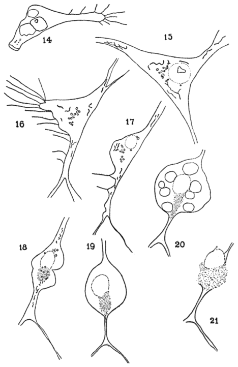
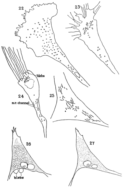
Hogue (1919) gave the word a definition and a picture, in a study not about movement. At the Carnegie Institution in Baltimore she grew explants of chick embryo heart at 39 °C in media of 0.225–1.8% NaCl (control 0.9%). The most concentrated medium, which draws water out of cells, killed established cultures within 3–4 h. One cell withdrew its processes and formed small marginal bulges, which her caption calls blebs; after 55 min, when it was recorded as dead, “a large bleb” surrounded its nucleus and cytoplasm. Of a second cell, whose attached parts had been drawn into streamers, she wrote: “blebs—clear, round cytoplasmic protrusions—appeared behind them” (Hogue, 1919, p. 634). This passing phrase is the definition.
What Hogue did not call blebs is as telling, for every use of the word concerns cells in hypertonic media. A recovering cell in 1.5% NaCl whose clear outer protoplasm, “resembling the ectoplasm of an ameba” (p. 635), formed blunt processes that retracted and re-formed elsewhere was not said to bleb. In our reading, a bleb was for Hogue a sign of a cell in trouble.
What it changed
For two decades after 1919 the word appears, among the papers we read, mostly in passing and for damaged cells, starting in Hogue’s own department (Lewis and Webster, 1921; Lewis, 1922). Reversible protrusions of healthy cells kept other names: Strangeways (1922, p. 140) saw “balloons of cytoplasm” on dividing chick cells, and workers on amoebae spoke of a ‘blister‘ when injected water lifted the membrane (Chambers and Reznikoff, 1926).
With phase-contrast microscopy and film the word returned to living cells. Zollinger (1948) found that suspended living cells of almost every type formed ‘blebs’ or ‘blisters’ within minutes, whereas dead cells never did. Costero and Pomerat (1951) filmed blebs forming and vanishing along dendrites of cultured neurons and named the process ‘zeiosis‘ (‘to boil over’). By the 1960s ‘bleb’ still competed with ‘bubbling’, ‘zeiosis’ and ‘potocytotic blistering’ (Price, 1967), and Prothero and Spencer (1968) found no systematic study of blebbing (M07).
In our reading the word thus covered from the start both reversible protrusions of living, moving cells, as for Ryder and Hardy, and signs of shrinkage and death, as for Hogue and her colleagues (proposed). As far as we found, none of the three cites another.
Later work and open questions
Where the word came from is unknown (open question). Ryder, Hardy and Hogue use it without attribution or claim to novelty, which in our reading suggests that it was already current in English; Hogue’s only cited source for such protrusions, Cash (1919), wrote of vesicles. Nor can these sources show whether the membrane had separated from a cortex, the criterion used since the late 1990s (M10).
A physical puzzle remains: a bleb needs an outward pressure, yet Hogue’s cells were losing water. Prothero and Spencer (1968) proposed that dying cells bleb because they take up water and raise their internal pressure, which fits Hogue’s shrinking cells only with added assumptions, such as the contraction she described (our reading). Both swelling and shrinkage can produce blebs: Cash (1919) saw vesicles in hypotonic saline and Zollinger (1948) saw blisters in distilled water and after hypertonic shrinkage, whereas Xenopus oocytes blebbed in hypertonic but not hypotonic medium (Zhang et al., 2000).
The ‘balloons‘ that grew from Hogue’s explants and floated free when their stalk snapped, which she did not call blebs, may have been shed blebs, an early record of the fragmentation described in M17. Zollinger (1948) read her as having seen blisters detach, but the drawings alone cannot decide.
References (16)
- Cash JR (1919) Reaction of cells in tissue culture to ether (abstract 13, Proc Am Assoc Anat). Anat Rec 16:146. doi:10.1002/ar.1090160304 (DOI of pp. 136–173).
- Chambers R, Reznikoff P (1926) Micrurgical studies in cell physiology. I. The action of the chlorides of Na, K, Ca, and Mg on the protoplasm of Amoeba proteus. J Gen Physiol 8:369–401. doi:10.1085/jgp.8.4.369
- Costero I, Pomerat CM (1951) Cultivation of neurons from the adult human cerebral and cerebellar cortex. Am J Anat 89:405–467. doi:10.1002/aja.1000890304
- Ecker A (1849) Zur Lehre vom Bau und Leben der contractilen Substanz der niedersten Thiere. Z Wiss Zool 1:218–245. No DOI.
- Hardy WB (1898) Further observations upon the action of the oxyphil and hyaline cells of frog’s lymph upon bacilli. J Physiol 23:359–375. doi:10.1113/jphysiol.1898.sp000733
- Hogue MJ (1919) The effect of hypotonic and hypertonic solutions on fibroblasts of the embryonic chick heart in vitro. J Exp Med 30:617–648. doi:10.1084/jem.30.6.617
- Kühne W (1864) Untersuchungen über das Protoplasma und die Contractilität. Leipzig: Engelmann. No DOI.
- Lewis MR (1922) The importance of dextrose in the medium of tissue cultures. J Exp Med 35:317–322. doi:10.1084/jem.35.3.317
- Lewis WH, Webster LT (1921) Wandering cells, endothelial cells, and fibroblasts in cultures from human lymph nodes. J Exp Med 34:397–405. doi:10.1084/jem.34.4.397
- Price ZH (1967) The micromorphology of zeiotic blebs in cultured human epithelial (HEp) cells. Exp Cell Res 48:82–92. doi:10.1016/0014-4827(67)90278-9
- Prothero JW, Spencer D (1968) A model of blebbing in mitotic tissue culture cells. Biophys J 8:41–51. doi:10.1016/S0006-3495(68)86473-2
- Ryder JA (1894) Dynamics in evolution. In: Biological Lectures delivered at the Marine Biological Laboratory of Wood’s Holl in the summer session of 1893. Boston: Ginn, pp. 63–81. No DOI.
- Strangeways TSP (1922) Observations on the changes seen in living cells during growth and division. Proc R Soc Lond B 94:137–141. doi:10.1098/rspb.1922.0050
- Verworn M (1892) Die Bewegung der lebendigen Substanz. Jena: Gustav Fischer. No DOI.
- Zhang Y, Gao F, Popov VL et al. (2000) Mechanically gated channel activity in cytoskeleton-deficient plasma membrane blebs and vesicles from Xenopus oocytes. J Physiol 523:117–130. doi:10.1111/j.1469-7793.2000.t01-1-00117.x
- Zollinger HU (1948) Cytologic studies with the phase microscope. I. The formation of “blisters” on cells in suspension (potocytosis), with observations on the nature of the cellular membrane. Am J Pathol 24:545–567. PMID 18859358