A long history of blebbing
How ideas about blebs changed over 270 years, from Rösel's shape-changing 'little Proteus' of 1755 through protoplasm, gels and pressure to the cortex and reconstituted blebs.
Our title nods to “A short history of blebbing”, in which Guillaume Charras told the life of a single bleb (Charras, 2008). A bleb is a rounded bulge of plasma membrane that forms where the membrane is no longer held by the cortex, a thin layer of actin filaments and myosin motors beneath it. It inflates for about 30 s, then rebuilds a cortex and is pulled back by myosin over about 2 min. Our history begins earlier, in 1755, and follows the older literature on amoeboid movement and on the cytoplasm as a gel, where several ideas now used for blebs first appeared.
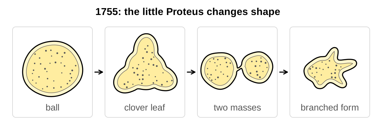
A little Proteus, 1755
Histories of amoeba research begin with a plate published by Rösel von Rosenhof in 1755 (Lorch, 1973; Reynolds, 2008). Rösel found tiny animals in standing river water that changed shape continually, and he called them the ‘little Proteus‘, after the shape-changing sea god. He drew one in sequence, from a ball and a clover leaf to two masses joined by a thin bridge and finally two animals: “Nun hatte ich statt eines Thieres zweye” (now I had two animals instead of one; Rösel von Rosenhof, 1755, p. 622).
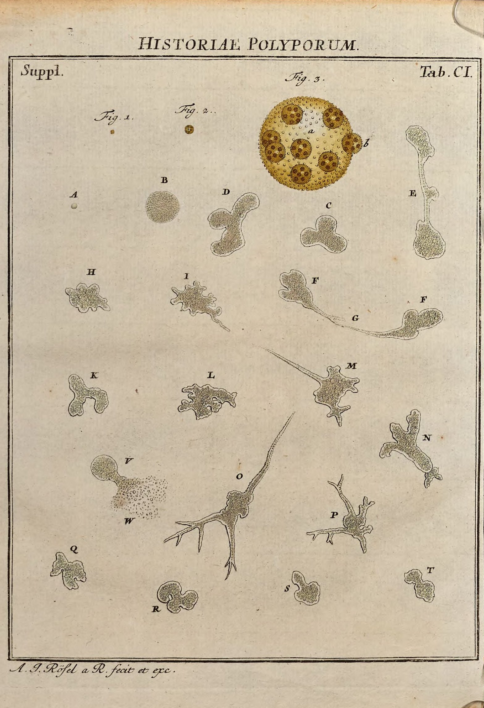
When Rösel held an animal fast with a quill, he found its granular substance “vermittelst eines zarten äusseren Häutleins zusammen gehalten” (held together by a delicate outer skin; Rösel von Rosenhof, 1755, p. 623), and when the skin tore, the animal flowed apart. Whether a cell’s surface is a separate layer, and what happens when it gives way, is a question this history keeps returning to (M01).
Protoplasm and the hernia, 1835–1865
The earliest mechanical explanation of amoeboid shape change that we found is Ehrenberg’s, although his 1830 memoir said only that amoebae change shape at will (Ehrenberg, 1830, p. 75). One spot of the body slackens, he later proposed, and contraction of the rest drives the contents into it, as into a hernia: “das ganze Geheimniss des Formenwechsels” (the whole secret of the change of form; Ehrenberg, 1838, p. 126). Dujardin (1835) saw no skin around amoebae and rejected the hernia, which needs a contractile envelope; in his view a protrusion of sarcode, his name for the living substance, advances by a force of its own. Whether a protrusion is pushed out from behind or advances by itself would be asked again of blebs. Ecker (1849) and Kühne (1864) sided with contraction, and theirs are the earliest accounts we found of a protrusion pushed out at a weak spot by contraction elsewhere, the scheme later applied to blebs (proposed).
Meanwhile colorless blood corpuscles were seen to crawl on a microscope stage heated to about 35 °C (Schultze, 1865). Schultze had redefined the cell as “ein Klümpchen Protoplasma, in dessen Innerem ein Kern liegt” (a lump of protoplasm with a nucleus inside; Schultze, 1861, p. 11), with the membrane inessential. Amoeboid movement thus became a property of protoplasm, and ‘amoeba’ came to name a behavior rather than an organism (Reynolds, 2008; M02).
The word ‘bleb’, 1893–1919
The word ‘bleb’ entered cell biology without ceremony. The earliest use we found is in an 1893 lecture by Ryder, who explained the shape of Amoeba proteus by the surface tension theory then in favor and noted “small blebs or pseudopodial warts” along a retracting pseudopod (Ryder, 1894, p. 76). Hardy (1898, p. 368) wrote of “little blebs” at the hind end of a moving frog lymph cell. The first definition we found comes from dying cells: as chick embryo fibroblasts shrank and died in concentrated saline, Hogue (1919, p. 634) saw “blebs—clear, round cytoplasmic protrusions” appear. The word thus had three early homes: a living amoeba, a moving white blood cell and dying cells. The split between blebs of moving cells and blebs of injured cells runs through the rest of this history. Earlier uses may exist, so which came first is an open question (M03).
A tube of gel around a fluid core, 1923–1947
As the surface tension theory lost ground, its replacement arrived twice in 1923. Mast (1923) described Amoeba proteus as a fluid core of plasmasol inside a solid layer of plasmagel (plasmagel and plasmasol), wrapped in a thin elastic membrane, with sol turning into gel at the front and gel dissolving at the rear. Pantin (1923, p. 31) described in marine amoebae “a contracting tube of gelated ectoplasm closed at its hind end”, through which the inner endoplasm flowed passively.
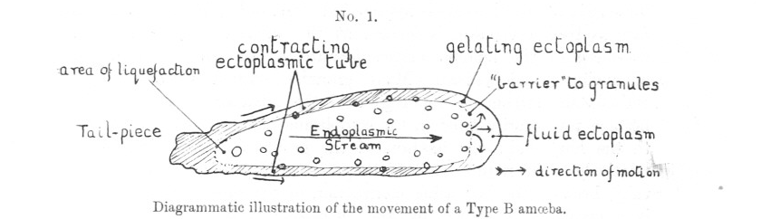
Mast’s scheme contained the parts of a bleb, although nobody called them that. The sol, he supposed, takes up water and presses outward, and at high temperature the gel at the tip of a pseudopod “often suddenly breaks” (Mast, 1923, p. 261): the sol rushes through and stretches the membrane into a bulge, and new gel forms beneath it. In our reading, this is the sequence of a bleb started by a tear in the cortex (proposed). Mast’s account was never tested, and the only time-resolved record we found shows the front layer of A. proteus detaching from the membrane about every 2 s rather than tearing (Grębecki, 1990). De Bruyn (1947) rejected Mast’s osmotic push but judged reversible sol–gel changes well established (M04).
Measuring pressure in giant amoebae, 1959–1961
For decades nobody measured the pressure that these schemes required. Allen and Roslansky (1959) let pseudopods of the giant amoeba Chaos chaos seal themselves into glass capillaries and found that a gas pressure of about 1 cm of water (≈108 Pa) stopped streaming. Yet suction of up to ≈590 Pa through one pseudopod had “virtually no effect” on others a few hundred micrometers away (Allen and Roslansky, 1959, p. 442). If flow was pressure-driven, the authors reasoned, the endoplasm had to be non-Newtonian, its viscosity changing with the rate of flow, and Yagi (1961) found that its viscosity fell about tenfold as streaming sped up, although he blamed changes of the colloid rather than shear. That the endoplasm is not a simple fluid is strongly supported. Whether pressure is uniform inside a moving cell returned as a question with blebs (M05).
Bubbling cells under the phase microscope, 1946–1968
From the late 1940s phase contrast, which shows living cells without staining, moved the study of protrusions to animal cells. Zollinger (1948) saw ‘blebs’ or ‘blisters’ form within minutes on living cells in suspension but never on dead ones, and Costero and Pomerat (1951) named the blebs that came and went on cultured neurons zeiosis, from a Greek word for boiling over. Filming dividing cells, Hughes (1952) saw blebs grow for seconds and shrink more slowly, credited the first sighting to Peremeschko (1879), and proposed that growth stops when the tension of the bleb wall balances the pressure of the cell.
Experiments soon made blebs on demand. Chick fibroblasts deprived of divalent cations by the chelator EDTA contracted and blebbed continuously, and the authors’ “admittedly speculative” explanation was that weakened patches of the surface were blown out by internal pressure (Dornfeld and Owczarzak, 1958, p. 248). Prothero and Spencer (1968) turned the blow-out into the earliest mathematical model of blebbing that we found. Using Laplace’s law, which links the pressure inside a curved surface to its tension and curvature, they argued that a membrane whose tension stays flat over a range of stretch bulges wherever pressure times radius exceeds that tension.
The assumed pressure was far too low. A membrane at their 10 pN/µm with a radius of 10 µm balances only about 1 Pa (our arithmetic), whereas later estimates reach 20–100 Pa (Dai and Sheetz, 1999; Tinevez et al., 2009), so the resisting tension had to move into the cortex. Their timing held better: blebs on dividing cells rose in about 6 s and lasted about 30 s (99 blebs on four cells), and fast growth with slower retraction was confirmed by later measurements (strongly supported; M07).
Contraction, and blebs that move cells, 1973–1984
In the early 1970s blebs moved away from injury. Cytochalasin D, an actin drug then thought to relax cells, instead made cultured cells contract within 2–3 min as knobs of inner cytoplasm appeared, a response that energy poisons blocked (Miranda et al., 1974a). In sections, filaments under the surface gathered into pads with cytoplasm bulging between them (Miranda et al., 1974b), a “herniation of endoplasm through undefended places of the cortex” (Godman et al., 1975, p. 644). Blebbing thus became an active result of contraction, in three steps that later work would test: contraction raises the pressure, the cortex opens, and cytoplasm herniates through the gap. The evidence came from one group and pressure was not measured, so the mechanism remained proposed (M08).
Embryologists meanwhile asked whether blebs move cells. Holtfreter (1946) had seen clear bulges travel around isolated amphibian embryonic cells without displacing them, a circus movement, while elongated cells crept forward when slight contact gave them “a sufficient frictional support” (Holtfreter, 1946, p. 43). In living Fundulus embryos, Trinkaus (1973) saw deep cells move at 2–30 µm/min behind blebs that led and steered them, and wrote “Blebs lead the way” (Trinkaus, 1973, p. 81). In collagen gels, lymphocytes pushed blebs into the matrix; the blebs stayed fixed while the cell body moved through a stationary constriction, which Haston and Shields (1984) likened to the peristalsis of worms. The idea rests on kinematics alone (M09).
The jelly becomes an actin network, 1979–2013
While cell biologists filmed blebs, biochemists took the gel apart. Pollard et al. (1982) recast the contractile jelly of protoplasm as a meshwork of actin filaments that gels above a critical concentration of cross-linker, and purified systems showed that such a gel must be connected to contract, but that too many connections stop it. Actomyosin linked by filamin contracted as a whole only above a threshold of cross-linking, yet more slowly as cross-linking rose further (Janson et al., 1991), and fascin-linked gels contracted into local clusters when weakly connected and into one body when strongly connected (Alvarado et al., 2013). Physicists call this behavior percolation: force crosses a network only once enough filaments are linked. The connectivity threshold for contraction is strongly supported in vitro.
Bleb research took this up late. In stable blebs, the large, persistent blebs with which some confined cells move, García-Arcos et al. (2024b ◆) measured an actin-occupancy threshold of 0.59 in HeLa cells and mapped the front, middle and rear of the bleb onto regimes of connectivity, a result so far from one cell line (M06).
Membrane freed from the cortex, 1992–1999
Actin probes then showed what a bleb is made of. In M2 melanoma cells, which lack the actin cross-linker filamin and bleb over their whole surface (Cunningham et al., 1992), Cunningham (1995) saw each bleb grow at a constant rate for 4–7 s and then stop abruptly. Phalloidin, which binds actin filaments, labeled blebs that had stopped growing but not those still expanding, so “a polymerized actin structure is not present initially” (Cunningham, 1995, p. 1589), and actin appears later and stops the expansion.
The decisive image came from Walker carcinosarcoma cells, a blebbing tumor line. Staining actin and membrane lipid, Keller and Eggli (1998, p. 183) saw the membrane bulge outward “whereas the actin layer stays in place”. A bleb could now be defined by its structure rather than its shape: membrane separated from the cortex, inflated by cytoplasm and only later lined with new actin (established). The freed membrane also served as an instrument: assuming that a tether pulled from a bleb reports membrane tension alone, Dai and Sheetz (1999) derived the membrane tension and, through Laplace’s law, an intracellular pressure of about 20 Pa in melanoma cells (M10).
One engine for dying and invading cells, 1998–2003
In the same years, the blebbing of dying cells, which by the early 2000s was the most common subject of bleb papers, was traced to its engine. In serum-deprived PC12 cells, an inhibitor of caspases, the proteases that execute apoptosis, prevented death but not blebbing, whereas inhibitors of myosin light-chain kinase reduced blebbing, and phosphorylation of the light chain, which activates myosin II, was three to five times higher in blebbing cultures (Mills et al., 1998). In apoptotic cells, caspases cut the kinase ROCK I into a form 7.8 ± 0.5 times more active (s.e.m.; Coleman et al., 2001), and inhibiting ROCK abolished light-chain phosphorylation and blebbing (Sebbagh et al., 2001).
The same engine drove tumor invasion. In Matrigel, a three-dimensional matrix, a ROCK inhibitor cut the invasion of rounded, blebbing A375m2 melanoma cells to 3.8% of control but spared elongated cells (Sahai and Marshall, 2003). By 2003 the blebs of dying and of invading cells thus shared myosin II activated by phosphorylation of its light chain (strongly supported). The routes upstream differed, however: blebbing of PC12 cells fell when Rho was inhibited but not when caspases were blocked, whereas the apoptotic blebbing studied in 2001 needed caspases but not Rho (M11).
Pressure, tension and the life of a bleb, 2005–2010
With structure and engine known, the questions became physical. When Charras et al. (2005) perfused drugs that lowered contraction or drew water out of the cell over one side of blebbing M2 cells, blebbing fell only on that side, which it should not have done had pressure equalized. They proposed that the cytoplasm is poroelastic, an elastic mesh soaked in fluid like a water-filled sponge, through which a change of pressure spreads over about 10 µm in about 10 s. Later measurements supported a poroelastic cytoplasm (Moeendarbary et al., 2013), which may also explain why suction failed to cross a giant amoeba in 1959 (our reading).
Tinevez et al. (2009) measured the other side of the balance. They cut a small hole in the cortex of suspended L929 fibroblasts with a laser while lowering cortical tension, measured by micropipette aspiration, in steps with a ROCK inhibitor; below about 200 pN/µm no bleb grew. Contraction of the cortex thus sets the pressure that drives a bleb, much as the rubber of a balloon pressurizes the air inside. The threshold is strongly supported, with its value measured so far in L929 cells alone.
The two studies read pressure differently. Tinevez et al. (2009) took the finding that a second bleb was about 25% smaller whether it formed near or far from the first as a sign of fast equalization, but a poroelastic model later concluded that “lack of equilibration is essential to explain the results from Tinevez” (Strychalski and Guy, 2016, p. 1174). M2 cells beside a micropipette that drained pressure kept blebbing, which Malboubi et al. (2026) read as pressure divided into compartments. The cells, perturbations and timescales differ, and how far and how fast pressure spreads in a blebbing cell remains open (M12).
How a bleb ends was described in the same years. At retracting blebs of M2 cells, the membrane–actin linker ezrin arrived first, as expansion stopped, then actin and cross-linkers, and myosin last, completing a contractile cortex in about 30 s (Charras et al., 2006). The coupling between membrane and cortex then became measurable: in the zebrafish embryo, lowering the function of the ERM proteins, which link membrane to actin, reduced the energy of membrane–cortex attachment and enlarged blebs (Diz-Muñoz et al., 2010; M13).
A way of migrating, 2006–2015
Blebs entered migration research through embryos and amoebae. Zebrafish primordial germ cells moving toward a chemokine advanced by protrusions that began where the membrane separated from the cortex and filled by cytoplasmic flow, and raising calcium at a germ cell’s rear made it bleb there (Blaser et al., 2006). Dictyostelium amoebae moving up a chemical gradient blebbed at their front, and cells lacking myosin II made other protrusions instead (Yoshida and Soldati, 2006). Blebs, driven by contraction rather than by actin polymerizing at the front, were thus a normal and steerable protrusion (established), although germ cells with reduced E-cadherin function blebbed but stalled (Kardash et al., 2010; M18).
Taking Trinkaus’s “Blebs lead the way” as their title, Charras and Paluch (2008) proposed that confined cells might bleb forward without specific adhesion. Under a roof 3–5 µm high on a non-adhesive surface, fibroblasts and 20 other cell types, normal epithelial cells excepted, did produce a fast subpopulation that moved with one large bleb at 5.3 ± 1.5 µm/min, against 0.234 ± 0.09 µm/min for slow mesenchymal cells (s.e.m.; Liu et al., 2015), and high contractility switched zebrafish progenitor cells to the same stable-bleb form (Ruprecht et al., 2015).
Cells migrating this way turned out to transmit force mainly by friction rather than by pushing against the walls. In channels whose coatings set the friction, Walker cells moved on high-friction surfaces but not on low-friction ones, their cortex flowing rearward and gripping the walls at stresses of about 1 Pa or less (Bergert et al., 2015). Whether cells use this mode in unperturbed tissue is less clear: the first spontaneous candidate, zebrafish T cells moving in a 3 µm gap between skin layers, was identified without a membrane or cortex marker (Robertson et al., 2025; M19, M20).
Blebs rebuilt, turned inside out and read as signals, 2016–2026
The last decade has asked what a bleb minimally needs and what else it does. Blebs were rebuilt inside liposomes from purified actin, myosin and a protein linking actin to the membrane (Loiseau et al., 2016). Such systems run at tensions far below those of cells, and none of their blebs has yet been seen to retract by rebuilding a cortex, but they can test whether a bleb starts where the cortex tears or where the membrane peels off an intact cortex. With slowly activated myosin, Miyazaki et al. (2026) obtained both routes, tearing becoming more common as actin–membrane coupling rose and less common as cross-linking rose.
In cells the route depends on the system. In nucleus-free L929 fragments a hole opened in the actin shell before a bulge grew (Paluch et al., 2005), whereas 88.7 ± 7.6% of blebs on Walker cells kept the old cortex at their base, as peeling predicts (mean ± s.d.; Keller et al., 2002). In Dictyostelium, one study saw the old cortex remain intact as a scar after each bleb formed (Zatulovskiy et al., 2014), while another found cortex gaps at initiation sites inferred from each bleb’s final shape (Asante-Asamani et al., 2022). The second approach is correlative, and the reason for the disagreement has not been tested. Which route migrating vertebrate cells use is an open question (M15).
Blebs also work in reverse. In zebrafish vessel sprouts, the luminal membrane bulged into endothelial cells as actin-free blebs that actomyosin then retracted; stopping blood flow stopped this inverse blebbing, and the lumen regressed (Gebala et al., 2016). Inward blebs in the 32-cell mouse embryo were proposed to pump fluid between small lumens, although there, as in the vessels, the pressure is inferred rather than measured (Schliffka et al., 2024; M14).
Finally, blebs were found to carry signals. In melanoma cells kept without adhesion, septins, proteins that recognize membrane curvature, gathered at the curved bases of blebs and built a signaling hub that kept ERK and PI3K signaling active, so that blebbing helped detached cells survive (Weems et al., 2023). An independent group found a septin–PI3K survival axis at the neck of drug-induced blebs (Zhen et al., 2025), but the test in tumors, still a preprint, perturbs septins rather than blebs (Weems et al., 2026 preprint; M16).
Pieces that leave the cell, 1889–2025
Rösel’s animal split in two, and pieces of cells have been seen to move on their own ever since. Anucleate pieces of amoebae kept moving (Hofer, 1889), and a HeLa bleb that had taken in the nucleus parted from its cell and moved for 11 h (Rose, 1966). Blebbing is not moving, however: fibroblast fragments made with cytochalasin blebbed for hours without moving from place to place (Albrecht-Buehler, 1980).
Recent work has shown that blebs are one route to such fragments. In confined HeLa cells, stable blebs pulled away until the membrane neck at their base broke, and the anucleate fragments, called cytokineplasts, moved for hours at 5–15 µm/min and crossed collagen (García-Arcos, 2020 ◆; García-Arcos et al., 2024b ◆). In mouse lungs, injected melanoma cells lodged in capillaries “began to shed microscale blebs” within 40 s (Headley et al., 2016, p. 513), and their anucleate fragments became most of the tumor material in the lung. Later papers, García-Arcos et al. (2024b ◆) among them, call these fragments bleb-derived, but only cytoplasmic and nuclear labels were imaged, so a bleb origin was not tested.
Detachment of blebs is strongly supported as one route to fragmentation, but how often it happens in tissue is unknown: of 195 papers on cell fragmentation that we mapped, only 19 image a bleb detaching, and none images the neck with membrane and cortex markers (M17).
What has lasted
In our reading, three ideas have held since the 19th century. Contraction of one part of a cell pushes out another, from Ehrenberg’s hernia and Pantin’s contracting tube to the cortical tension that pressurizes a blebbing cell. The boundary between membrane and cortex decides where a bleb forms, whether by tearing or by peeling. And cells shed pieces that keep moving. What remains open is mostly about tissue: whether blebs are needed there rather than merely present, how blebbing cells transmit force, and whether the pressure inside them is uniform or local.
References (75)
- Albrecht-Buehler G (1980) Autonomous movements of cytoplasmic fragments. Proc Natl Acad Sci USA 77:6639–6643. doi:10.1073/pnas.77.11.6639
- Allen RD, Roslansky JD (1959) The consistency of ameba cytoplasm and its bearing on the mechanism of ameboid movement. I. An analysis of endoplasmic velocity profiles of Chaos chaos (L.). J Biophys Biochem Cytol 6:437–446. doi:10.1083/jcb.6.3.437
- Alvarado J, Sheinman M, Sharma A et al. (2013) Molecular motors robustly drive active gels to a critically connected state. Nat Phys 9:591–597. doi:10.1038/nphys2715
- Asante-Asamani E, Grange D, Rawal D et al. (2022) A role for myosin II clusters and membrane energy in cortex rupture for Dictyostelium discoideum. PLoS ONE e0265380. doi:10.1371/journal.pone.0265380
- Bergert M, Erzberger A, Desai RA et al. (2015) Force transmission during adhesion-independent migration. Nat Cell Biol 17:524–529. doi:10.1038/ncb3134
- Blaser H, Reichman-Fried M, Castanon I et al. (2006) Migration of zebrafish primordial germ cells: a role for myosin contraction and cytoplasmic flow. Dev Cell 11:613–627. doi:10.1016/j.devcel.2006.09.023
- Charras GT, Yarrow JC, Horton MA et al. (2005) Non-equilibration of hydrostatic pressure in blebbing cells. Nature 435:365–369. doi:10.1038/nature03550
- Charras GT, Hu CK, Coughlin M et al. (2006) Reassembly of contractile actin cortex in cell blebs. J Cell Biol 175:477–490. doi:10.1083/jcb.200602085
- Charras GT (2008) A short history of blebbing. J Microsc 231:466–478. doi:10.1111/j.1365-2818.2008.02059.x
- Charras G, Paluch E (2008) Blebs lead the way: how to migrate without lamellipodia. Nat Rev Mol Cell Biol 9:730–736. doi:10.1038/nrm2453
- Coleman ML, Sahai EA, Yeo M et al. (2001) Membrane blebbing during apoptosis results from caspase-mediated activation of ROCK I. Nat Cell Biol 3:339–345. doi:10.1038/35070009
- Costero I, Pomerat CM (1951) Cultivation of neurons from the adult human cerebral and cerebellar cortex. Am J Anat 89:405–467. doi:10.1002/aja.1000890304
- Cunningham CC, Gorlin JB, Kwiatkowski DJ et al. (1992) Actin-binding protein requirement for cortical stability and efficient locomotion. Science 255:325–327. doi:10.1126/science.1549777
- Cunningham CC (1995) Actin polymerization and intracellular solvent flow in cell surface blebbing. J Cell Biol 129:1589–1599. doi:10.1083/jcb.129.6.1589
- Dai J, Sheetz MP (1999) Membrane tether formation from blebbing cells. Biophys J 77:3363–3370. doi:10.1016/S0006-3495(99)77168-7
- De Bruyn PPH (1947) Theories of amoeboid movement. Q Rev Biol 22:1–24. doi:10.1086/395577
- Diz-Muñoz A, Krieg M, Bergert M et al. (2010) Control of directed cell migration in vivo by membrane-to-cortex attachment. PLoS Biol 8:e1000544. doi:10.1371/journal.pbio.1000544
- Dornfeld EJ, Owczarzak A (1958) Surface responses in cultured fibroblasts elicited by ethylenediaminetetraacetic acid. J Biophys Biochem Cytol 4:243–250. doi:10.1083/jcb.4.3.243
- Dujardin F (1835) Recherches sur les organismes inférieurs. Ann Sci Nat Zool (2e sér.) 4:343–377. No DOI.
- Ecker A (1849) Zur Lehre vom Bau und Leben der contractilen Substanz der niedersten Thiere. Z Wiss Zool 1:218–245. No DOI.
- Ehrenberg CG (1830) Beiträge zur Kenntniss der Organisation der Infusorien und ihrer geographischen Verbreitung, besonders in Sibirien. Abh Akad Wiss Berlin 1830 (Phys Abh):1–88, Taf. I–VIII. doi:10.5962/bhl.title.143632
- Ehrenberg CG (1838) Die Infusionsthierchen als vollkommene Organismen. Leipzig: Voss. pp. 125–129. No DOI.
- ◆ García-Arcos JM (2020) Stabilization and motility mechanism of blebs in cancer cells. PhD thesis, Université de Paris (Institut Curie). doi:10.70675/a35575c7z85b5z4ad1z98acz50f8d65ebbac
- ◆ García-Arcos JM, Ziegler J, Grigolon S et al. (2024b) Rigidity percolation and active advection synergize in the actomyosin cortex to drive amoeboid cell motility. Dev Cell 59:2990–3007.e7. doi:10.1016/j.devcel.2024.06.023
- Gebala V, Collins R, Geudens I et al. (2016) Blood flow drives lumen formation by inverse membrane blebbing during angiogenesis in vivo. Nat Cell Biol 18:443–450. doi:10.1038/ncb3320
- Godman GC, Miranda AF, Deitch AD et al. (1975) Action of cytochalasin D on cells of established lines. III. Zeiosis and movements at the cell surface. J Cell Biol 64:644–667. doi:10.1083/jcb.64.3.644
- Grębecki A (1990) Dynamics of the contractile system in the pseudopodial tips of normally locomoting amoebae, demonstrated in vivo by video-enhancement. Protoplasma 154:98–111. doi:10.1007/BF01539837
- Hardy WB (1898) Further observations upon the action of the oxyphil and hyaline cells of frog’s lymph upon bacilli. J Physiol 23:359–375. doi:10.1113/jphysiol.1898.sp000733
- Haston WS, Shields JM (1984) Contraction waves in lymphocyte locomotion. J Cell Sci 68:227–241. doi:10.1242/jcs.68.1.227
- Headley MB, Bins A, Nip A et al. (2016) Visualization of immediate immune responses to pioneer metastatic cells in the lung. Nature 531:513–517. doi:10.1038/nature16985
- Hofer B (1889) Experimentelle Untersuchungen über den Einfluss des Kerns auf das Protoplasma. Jena Z Naturwiss 24:105–176. No DOI.
- Hogue MJ (1919) The effect of hypotonic and hypertonic solutions on fibroblasts of the embryonic chick heart in vitro. J Exp Med 30:617–648. doi:10.1084/jem.30.6.617
- Holtfreter J (1946) Structure, motility and locomotion in isolated embryonic amphibian cells. J Morphol 79:27–62. doi:10.1002/jmor.1050790103
- Hughes A (1952) The Mitotic Cycle: the Cytoplasm and Nucleus during Interphase and Mitosis. Butterworths, London. No DOI.
- Janson LW, Kolega J, Taylor DL (1991) Modulation of contraction by gelation/solation in a reconstituted motile model. J Cell Biol 114:1005–1015. doi:10.1083/jcb.114.5.1005
- Kardash E, Reichman-Fried M, Maître JL et al. (2010) A role for Rho GTPases and cell–cell adhesion in single-cell motility in vivo. Nat Cell Biol 12:47–53. doi:10.1038/ncb2003
- Keller H, Eggli P (1998) Protrusive activity, cytoplasmic compartmentalization, and restriction rings in locomoting blebbing Walker carcinosarcoma cells are related to detachment of cortical actin from the plasma membrane. Cell Motil Cytoskeleton 41:181–193. doi:10.1002/(SICI)1097-0169(1998)41:2<181::AID-CM8>3.0.CO;2-H
- Keller H, Rentsch P, Hagmann J (2002) Differences in cortical actin structure and dynamics document that different types of blebs are formed by distinct mechanisms. Exp Cell Res 277:161–172. doi:10.1006/excr.2002.5552
- Kühne W (1864) Untersuchungen über das Protoplasma und die Contractilität. Leipzig: Engelmann. No DOI.
- Liu YJ, Le Berre M, Lautenschlaeger F et al. (2015) Confinement and low adhesion induce fast amoeboid migration of slow mesenchymal cells. Cell 160:659–672. doi:10.1016/j.cell.2015.01.007
- Loiseau E, Schneider JAM, Keber FC et al. (2016) Shape remodeling and blebbing of active cytoskeletal vesicles. Sci Adv 2:e1500465. doi:10.1126/sciadv.1500465
- Lorch IJ (1973) Some historical aspects of amoeba studies. In: Jeon KW (ed.) The Biology of Amoeba. New York: Academic Press, pp. 1–36 (pp. 1–14 read). No DOI.
- Malboubi M, Esteki MH, Vaghela MB et al. (2026) The cytoplasm of living cells can sustain transient and steady intracellular pressure gradients. eLife 14:RP105523. doi:10.7554/eLife.105523
- Mast SO (1923) Mechanics of locomotion in Amoeba. Proc Natl Acad Sci USA 9:258–261. doi:10.1073/pnas.9.7.258
- Mills JC, Stone NL, Erhardt J et al. (1998) Apoptotic membrane blebbing is regulated by myosin light chain phosphorylation. J Cell Biol 140:627–636. doi:10.1083/jcb.140.3.627
- Miranda AF, Godman GC, Deitch AD et al. (1974a) Action of cytochalasin D on cells of established lines. I. Early events. J Cell Biol 61:481–500. doi:10.1083/jcb.61.2.481
- Miranda AF, Godman GC, Tanenbaum SW (1974b) Action of cytochalasin D on cells of established lines. II. Cortex and microfilaments. J Cell Biol 62:406–423. doi:10.1083/jcb.62.2.406
- Miyazaki M, Laboni FS, Kim T (2026) Reconstitution of actomyosin networks in cell-sized liposomes dissects distinct mechanisms of membrane blebbing and symmetry breaking. Sci Adv 12:eaed8818. doi:10.1126/sciadv.aed8818
- Moeendarbary E, Valon L, Fritzsche M et al. (2013) The cytoplasm of living cells behaves as a poroelastic material. Nat Mater 12:253–261. doi:10.1038/nmat3517
- Paluch E, Piel M, Prost J et al. (2005) Cortical actomyosin breakage triggers shape oscillations in cells and cell fragments. Biophys J 89:724–733. doi:10.1529/biophysj.105.060590
- Pantin CFA (1923) On the physiology of amoeboid movement. I. J Mar Biol Assoc UK 13:24–69. doi:10.1017/S0025315400010924
- Peremeschko (1879) Ueber die Theilung der thierischen Zellen. Arch Mikrosk Anat 16:437–457. doi:10.1007/bf02956388
- Pollard TD, Aebi U, Cooper JA et al. (1982) Actin structure, polymerization, and gelation. Cold Spring Harb Symp Quant Biol 46:513–524. doi:10.1101/SQB.1982.046.01.048
- Prothero JW, Spencer D (1968) A model of blebbing in mitotic tissue culture cells. Biophys J 8:41–51. doi:10.1016/S0006-3495(68)86473-2
- Reynolds A (2008) Amoebae as exemplary cells: the protean nature of an elementary organism. J Hist Biol 41:307–337. doi:10.1007/s10739-007-9142-8
- Robertson TF, Schrope J, Zwick Z et al. (2025) Live imaging in zebrafish reveals tissue-specific strategies for amoeboid migration. Development 152:dev204351. doi:10.1242/dev.204351 (read as bioRxiv doi:10.1101/2024.08.14.607647)
- Rose GG (1966) Zeiosis. I. Ejection of cell nuclei into zeiotic blebs. J R Microsc Soc 86:87–102. doi:10.1111/j.1365-2818.1966.tb05328.x
- Rösel von Rosenhof AJ (1755) Der monatlich-herausgegebenen Insecten-Belustigung Dritter Theil. Nürnberg, pp. 615–624, Tab. CI. No DOI.
- Ruprecht V, Wieser S, Callan-Jones A et al. (2015) Cortical contractility triggers a stochastic switch to fast amoeboid cell motility. Cell 160:673–685. doi:10.1016/j.cell.2015.01.008
- Ryder JA (1894) Dynamics in evolution. In: Biological Lectures delivered at the Marine Biological Laboratory of Wood’s Holl in the summer session of 1893. Boston: Ginn, pp. 63–81. No DOI.
- Sahai E, Marshall CJ (2003) Differing modes of tumour cell invasion have distinct requirements for Rho/ROCK signalling and extracellular proteolysis. Nat Cell Biol 5:711–719. doi:10.1038/ncb1019
- Schliffka MF, Dumortier JG, Pelzer D et al. (2024) Inverse blebs operate as hydraulic pumps during mouse blastocyst formation. Nat Cell Biol 26:1669–1677. doi:10.1038/s41556-024-01501-z
- Schultze M (1861) Ueber Muskelkörperchen und das, was man eine Zelle zu nennen habe. Arch Anat Physiol Wiss Med 1861:1–27. No DOI.
- Schultze M (1865) Ein heizbarer Objecttisch und seine Verwendung bei Untersuchungen des Blutes. Arch Mikrosk Anat 1:1–42. No DOI.
- Sebbagh M, Renvoizé C, Hamelin J et al. (2001) Caspase-3-mediated cleavage of ROCK I induces MLC phosphorylation and apoptotic membrane blebbing. Nat Cell Biol 3:346–352. doi:10.1038/35070019
- Strychalski W, Guy RD (2016) Intracellular pressure dynamics in blebbing cells. Biophys J 110:1168–1179. doi:10.1016/j.bpj.2016.01.012
- Tinevez JY, Schulze U, Salbreux G et al. (2009) Role of cortical tension in bleb growth. Proc Natl Acad Sci USA 106:18581–18586. doi:10.1073/pnas.0903353106
- Trinkaus JP (1973) Surface activity and locomotion of Fundulus deep cells during blastula and gastrula stages. Dev Biol 30:68–103. doi:10.1016/0012-1606(73)90049-3
- Weems AD, Welf ES, Driscoll MK et al. (2023) Blebs promote cell survival by assembling oncogenic signalling hubs. Nature 615:517–525. doi:10.1038/s41586-023-05758-6
- Weems AD, Islam MT, Borges H et al. (2026) In vivo evidence for bleb-induced survival signaling in metastatic melanoma. bioRxiv. doi:10.64898/2026.01.05.697601 preprint
- Yagi K (1961) The mechanical and colloidal properties of Amoeba protoplasm and their relations to the mechanism of amoeboid movement. Comp Biochem Physiol 3:73–91. doi:10.1016/0010-406X(61)90134-7
- Yoshida K, Soldati T (2006) Dissection of amoeboid movement into two mechanically distinct modes. J Cell Sci 119:3833–3844. doi:10.1242/jcs.03152
- Zatulovskiy E, Tyson R, Bretschneider T et al. (2014) Bleb-driven chemotaxis of Dictyostelium cells. J Cell Biol 204:1027–1044. doi:10.1083/jcb.201306147
- Zhen Z, Zhang C, Li J et al. (2025) Blebs regulate phosphoinositide distribution and promote cell survival through the Septin-SH3KBP1-PI3K axis. Am J Physiol Cell Physiol 329:C145–C158. doi:10.1152/ajpcell.00096.2025
- Zollinger HU (1948) Cytologic studies with the phase microscope. I. The formation of “blisters” on cells in suspension (potocytosis), with observations on the nature of the cellular membrane. Am J Pathol 24:545–567. PMID 18859358