Kinds of blebs
All blebs share one physical recipe, but they differ in how long they last and how they behave: transient, circus and stable blebs, leader blebs, and blebs of dividing, dying and squeezed cells.
Every bleb starts in the same way: the membrane loses the support of the cortex, the thin layer of actin and myosin beneath it, and the pressure inside the cell pushes it outward. What follows varies. Some blebs are pulled back within a minute, some travel around the cell like a wave, and some never retract and instead lead a cell that migrates for hours. The shared mechanism is explained in Physics of blebs.
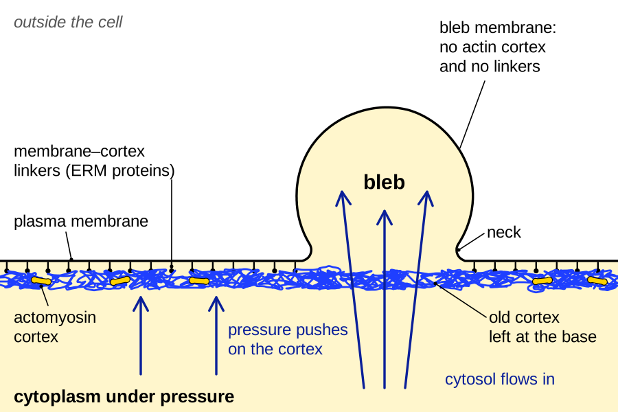
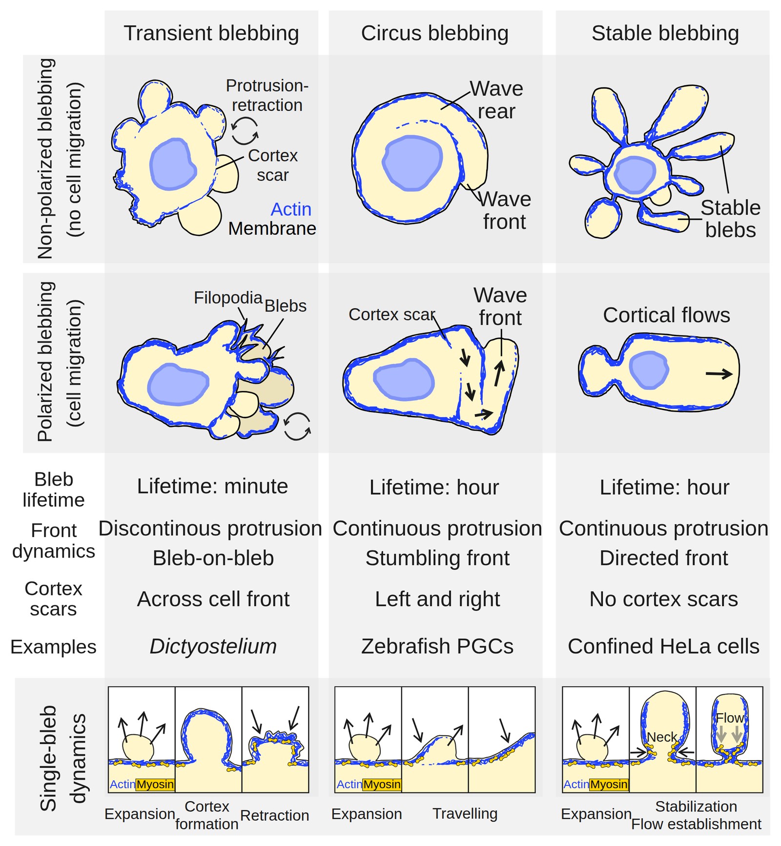
Sorting blebs by lifetime and by how their front moves gives three “blebbing regimes“: transient, circus and stable (García-Arcos et al., 2024a ◆). The grouping is descriptive rather than the result of a systematic comparison, and one cell can switch regimes.
Transient blebs: the classic cycle
The textbook bleb is transient: it inflates in seconds, stops, acquires a new cortex and is pulled back, a bleb cycle of about a minute. On M2 melanoma cells, expansion lasted 4–7 s, the bleb then stayed still for 10–20 s, and retraction took 40–60 s (Cunningham, 1995). Retraction is slower because it must wait until a contractile cortex has been rebuilt under the bleb membrane, which in the same cells took about 30 s (Charras et al., 2006).
Because each bleb rebuilds its own cortex, the old cortex remains as a “cortex scar” at the base of the new bleb (García-Arcos et al., 2024a ◆), and the surface advances in steps. If new blebs form preferentially on one side, the steps add up to migration. Dictyostelium amoebae chemotaxing in buffer or under agarose move in this way, forming their blebs mostly at the front, facing the source of the attractant (Yoshida and Soldati, 2006; Zatulovskiy et al., 2014).
Zebrafish primordial germ cells, which migrate through the embryo to the future gonad, bleb in the same way during their “run” phases (Blaser et al., 2006). Most protrusions at their front are blebs (84%; Olguin-Olguin et al., 2021), formed about once or twice a minute (Goudarzi et al., 2017; Olguin-Olguin et al., 2021). Each grows from a patch of actin at the leading edge, the “brush”: inflowing cytoplasm carries the front beyond it, and a new brush forms at the new front (Kardash et al., 2010). Between runs the cells lose their polarity and protrude in all directions, a phase called tumbling (Reichman-Fried et al., 2004), and isolated from the embryo they bleb extensively but without direction (Kardash et al., 2010).
Circus blebs: a wave around the cell
A circus bleb does not retract where it formed but travels around the cell edge. This “circus movement” was first described in dissociated embryonic cells. In cells from Xenopus gastrulae, a hyaline bleb rotated around the cell, clockwise or counterclockwise, at 0.9 ± 0.2 µm/s (mean ± s.d.), about 54 µm/min, and stopped wherever cells touched (Johnson, 1976). Medaka fish cells behaved similarly (Fujinami and Kageyama, 1975 abstract only), and waves of up to 90 µm/min have since been described along the cell contour (García-Arcos et al., 2024a ◆). Zebrafish primordial germ cells, which make polarized transient blebs in the embryo, mostly form circus blebs once dissociated from it (Goudarzi et al., 2017).
Electron microscopy of medaka cells gave the first structural clue: actin-like material lay in the cortex around the cell but was almost absent under the membrane at the front of the moving bleb (Fujinami, 1976 abstract only). Traveling blebs of dividing HeLa cells show this asymmetry directly, with a cortex-free front, contracting actomyosin at the rear pushing the cytosol forward, and a constant bleb volume (Charras, 2008). The wave is thus an asymmetric bleb cycle, the membrane detaching at its front while a cortex re-forms and contracts at its rear (García-Arcos et al., 2024a ◆).
A circus bleb spins around an isolated round cell without moving it, but a wave confined to one side can move the cell, as Johnson (1976) already saw. Neutrophil-like HL-60 cells lacking the branched-actin regulator WAVE advanced in this way under strong confinement: their blebs ran back and forth along a leading edge, often reversing, in a pattern the authors compared with circus movement, within a zone marked by Rac activity (Graziano et al., 2019). How common such a swinging front is among migrating cells is an open question.
Stable blebs and leader blebs
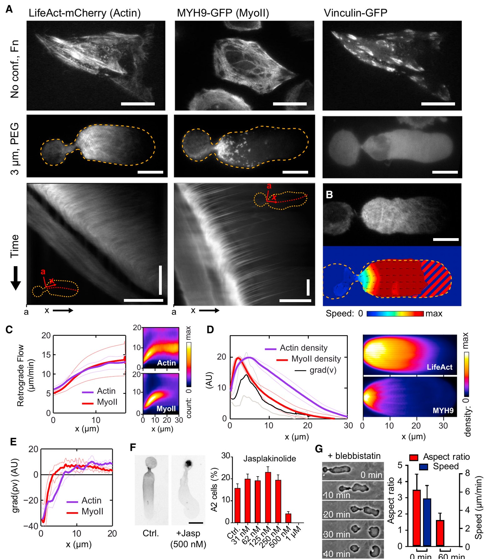
At the other extreme, a stable bleb never retracts: it is large, often elongated, can hold most of the cytoplasm, and persists for hours in cells that are strongly contractile, or squeezed and deprived of adhesion. Under a roof a few micrometers high on a non-adhesive surface, a fraction of human fibroblasts and of many other cell types switched to fast migration by forming a large bleb into which most of the cytoplasm and the nucleus moved, the elongated “A2” mode (Liu et al., 2015). Zebrafish progenitor cells made more contractile by serum, lysophosphatidic acid or confinement made the same switch stochastically (Ruprecht et al., 2015). In confined melanoma and other cancer cells, the large bleb is joined to the cell body by a narrow neck and leads the cell, hence the name “leader bleb” (Logue et al., 2015). The three terms now describe closely related behaviors (A1 and A2 modes).
A stable bleb does not retract because its cortex flows continuously rearward: actin and myosin assemble near the front, flow back toward the neck and are taken apart at the rear. The flow reached up to 150 µm/min at the very front in progenitor cells (Ruprecht et al., 2015) and about 16 µm/min in A2 cells (Liu et al., 2015). Because this cortical flow carries the network away from the front as fast as it forms, no complete, contracting cortex covers the tip, and nothing pulls it back (García-Arcos et al., 2024a ◆). The same flow, coupled to the surroundings by friction, propels the cell (Bergert et al., 2015). In confined HeLa cells, stable blebs persisted for several hours, and some detached as motile fragments (García-Arcos et al., 2024b ◆; see Fragments).
What makes the regimes different
One proposal is that the three regimes differ in how much the cortex re-forms under the membrane at the front, and when (García-Arcos et al., 2024a ◆). In a transient bleb the cortex re-forms completely before contraction begins, so the whole bleb is pulled back; in a circus bleb it re-forms on one side only, so the bleb moves along the surface; and in a stable bleb the rearward flow starts before the cortex covers the tip, which keeps protruding.
Work on confined HeLa cells gives this proposal a physical form. Transient blebs stopped growing when the new cortex covered their tip and retracted once actomyosin began to flow backward, whereas in blebs that became stable the flow started first (García-Arcos, 2020 ◆; García-Arcos et al., 2024b ◆, Fig. S7). In a model in which myosin carries actin filaments rearward and the network connects mechanically only above a threshold density, fast contraction cleared actin from the front and stabilized the bleb, whereas a bleb that actin filled first was retracted (García-Arcos et al., 2024b ◆). More contractility could thus stabilize blebs rather than retract them (proposed; see Physics of blebs).
Cells move between regimes as their conditions change. Dictyostelium cells compressed under a load switched from pseudopods to blebs within about 10 s, half of them at about 100 Pa, a switch that required the mechanosensitive channel Piezo and external calcium (Srivastava et al., 2020). Other cells seem to use other sensors for confinement, so there may be no single switch common to all (Migration).
Blebs in dividing, dying and squeezed cells
Other blebs are defined by the state of the cell rather than by their dynamics. Dividing cells bleb at their poles during cytokinesis; in L929 cells, blocking these blebs made the cleavage furrow oscillate and division fail in a fraction of cells, and the authors proposed that polar blebs act as valves that release cortical contractility (Sedzinski et al., 2011). Cells dying by apoptosis bleb vigorously, driven by actomyosin contraction (Mills et al., 1998) that, in the cell lines studied, is switched on when caspase-3 cleaves and activates the kinase ROCK I (Coleman et al., 2001). These apoptotic blebs are the subject of Dying and injured cells.
Where the pressure outside a cell is higher than inside, as in the lumen of a growing blood vessel or the fluid-filled cavity of an early mouse embryo, the membrane bulges into the cell without actin and is then pulled back by actomyosin, forming inverse blebs (Gebala et al., 2016; Schliffka et al., 2024; see M14). A bleb can also leave the cell altogether, carrying cytoplasm and sometimes the nucleus, which links blebbing to cell fragmentation (M17).
Things that look like blebs
A rounded bulge is not necessarily a bleb, and shape alone does not reveal how a protrusion formed. Lobopodia of fibroblasts in three-dimensional matrix are blunt protrusions thought to be driven by pressure, but they depend on integrin adhesions to the matrix, unlike the blebs of rounded cancer cells (Petrie et al., 2012). Blebbisomes are large extracellular vesicles that themselves bleb but do not arise from a bleb of the parent cell (Jeppesen et al., 2025), and blebs of the nuclear envelope are a different structure, with their own page. We call a protrusion a bleb when there is evidence that membrane grew free of the cortex under pressure, and say so where that evidence is missing.
How these kinds of blebs were recognized is told in A long history of blebbing, and the Milestones describe the key experiments.
References (29)
- Bergert M, Erzberger A, Desai RA et al. (2015) Force transmission during adhesion-independent migration. Nat Cell Biol 17:524–529. doi:10.1038/ncb3134
- Blaser H, Reichman-Fried M, Castanon I et al. (2006) Migration of zebrafish primordial germ cells: a role for myosin contraction and cytoplasmic flow. Dev Cell 11:613–627. doi:10.1016/j.devcel.2006.09.023
- Charras GT (2008) A short history of blebbing. J Microsc 231:466–478. doi:10.1111/j.1365-2818.2008.02059.x
- Charras GT, Hu CK, Coughlin M et al. (2006) Reassembly of contractile actin cortex in cell blebs. J Cell Biol 175:477–490. doi:10.1083/jcb.200602085
- Coleman ML, Sahai EA, Yeo M et al. (2001) Membrane blebbing during apoptosis results from caspase-mediated activation of ROCK I. Nat Cell Biol 3:339–345. doi:10.1038/35070009
- Cunningham CC (1995) Actin polymerization and intracellular solvent flow in cell surface blebbing. J Cell Biol 129:1589–1599. doi:10.1083/jcb.129.6.1589
- Fujinami N, Kageyama T (1975) Circus movement in dissociated embryonic cells of a teleost, Oryzias latipes. J Cell Sci 19:169–182. doi:10.1242/jcs.19.1.169 abstract only
- Fujinami N (1976) Studies on the mechanism of circus movement in dissociated embryonic cells of a teleost, Oryzias latipes: fine-structural observations. J Cell Sci 22:133–147. doi:10.1242/jcs.22.1.133 abstract only
- ◆ García-Arcos JM, Jha A, Waterman CM et al. (2024a) Blebology: principles of bleb-based migration. Trends Cell Biol 34:838–853. doi:10.1016/j.tcb.2024.02.009
- ◆ García-Arcos JM (2020) Stabilization and motility mechanism of blebs in cancer cells. PhD thesis, Université de Paris (Institut Curie). doi:10.70675/a35575c7z85b5z4ad1z98acz50f8d65ebbac
- ◆ García-Arcos JM, Ziegler J, Grigolon S et al. (2024b) Rigidity percolation and active advection synergize in the actomyosin cortex to drive amoeboid cell motility. Dev Cell 59:2990–3007.e7. doi:10.1016/j.devcel.2024.06.023
- Gebala V, Collins R, Geudens I et al. (2016) Blood flow drives lumen formation by inverse membrane blebbing during angiogenesis in vivo. Nat Cell Biol 18:443–450. doi:10.1038/ncb3320
- Goudarzi M, Tarbashevich K, Mildner K et al. (2017) Bleb expansion in migrating cells depends on supply of membrane from cell surface invaginations. Dev Cell 43:577–587.e5. doi:10.1016/j.devcel.2017.10.030
- Graziano BR, Town JP, Sitarska E et al. (2019) Cell confinement reveals a branched-actin independent circuit for neutrophil polarity. PLoS Biol 17:e3000457. doi:10.1371/journal.pbio.3000457
- Jeppesen DK, Sanchez ZC, Kelley NM et al. (2025) Blebbisomes are large, organelle-rich extracellular vesicles with cell-like properties. Nat Cell Biol 27:438–448. doi:10.1038/s41556-025-01621-0
- Johnson KE (1976) Circus movements and blebbing locomotion in dissociated embryonic cells of an amphibian, Xenopus laevis. J Cell Sci 22:575–583. doi:10.1242/jcs.22.3.575
- Kardash E, Reichman-Fried M, Maître JL et al. (2010) A role for Rho GTPases and cell–cell adhesion in single-cell motility in vivo. Nat Cell Biol 12:47–53. doi:10.1038/ncb2003
- Liu YJ, Le Berre M, Lautenschlaeger F et al. (2015) Confinement and low adhesion induce fast amoeboid migration of slow mesenchymal cells. Cell 160:659–672. doi:10.1016/j.cell.2015.01.007
- Logue JS, Cartagena-Rivera AX, Baird MA et al. (2015) Erk regulation of actin capping and bundling by Eps8 promotes cortex tension and leader bleb-based migration. eLife 4:e08314. doi:10.7554/eLife.08314
- Mills JC, Stone NL, Erhardt J et al. (1998) Apoptotic membrane blebbing is regulated by myosin light chain phosphorylation. J Cell Biol 140:627–636. doi:10.1083/jcb.140.3.627
- Olguin-Olguin A, Aalto A, Maugis B et al. (2021) Chemokine-biased robust self-organizing polarization of migrating cells in vivo. Proc Natl Acad Sci USA 118:e2018480118. doi:10.1073/pnas.2018480118
- Petrie RJ, Gavara N, Chadwick RS, Yamada KM (2012) Nonpolarized signaling reveals two distinct modes of 3D cell migration. J Cell Biol 197:439–455. doi:10.1083/jcb.201201124
- Reichman-Fried M, Minina S, Raz E (2004) Autonomous modes of behavior in primordial germ cell migration. Dev Cell 6:589–596. doi:10.1016/s1534-5807(04)00074-7
- Ruprecht V, Wieser S, Callan-Jones A et al. (2015) Cortical contractility triggers a stochastic switch to fast amoeboid cell motility. Cell 160:673–685. doi:10.1016/j.cell.2015.01.008
- Schliffka MF, Dumortier JG, Pelzer D et al. (2024) Inverse blebs operate as hydraulic pumps during mouse blastocyst formation. Nat Cell Biol 26:1669–1677. doi:10.1038/s41556-024-01501-z
- Sedzinski J, Biro M, Oswald A et al. (2011) Polar actomyosin contractility destabilizes the position of the cytokinetic furrow. Nature 476:462–466. doi:10.1038/nature10286
- Srivastava N, Traynor D, Piel M et al. (2020) Pressure sensing through Piezo channels controls whether cells migrate with blebs or pseudopods. Proc Natl Acad Sci USA 117:2506–2512. doi:10.1073/pnas.1905730117
- Yoshida K, Soldati T (2006) Dissection of amoeboid movement into two mechanically distinct modes. J Cell Sci 119:3833–3844. doi:10.1242/jcs.03152
- Zatulovskiy E, Tyson R, Bretschneider T et al. (2014) Bleb-driven chemotaxis of Dictyostelium cells. J Cell Biol 204:1027–1044. doi:10.1083/jcb.201306147