Fragments: when blebs leave the cell
How blebs can detach to become motile fragments or vesicles, how strong the evidence is, and how cytoplasts, apoptotic bodies, platelets, large oncosomes and blebbisomes differ.
A bleb is a rounded protrusion of the plasma membrane, pushed out by the pressure of the cytoplasm where the cortex, the thin network of actin filaments and myosin motors under the membrane, is missing or has come away. Most blebs are short-lived: a new cortex assembles under the bleb membrane, contracts and draws the bleb back. Sometimes, however, a bleb moves away from the cell body on a narrowing neck, the neck parts, and the freed piece of cytoplasm, usually without a nucleus, goes on blebbing, crawls on its own or carries material to other cells (Rose, 1966; Tadokoro et al., 2016; García-Arcos et al., 2024b ◆).
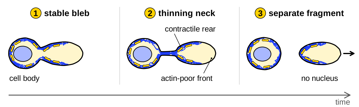
Bleb detachment has been imaged in several systems, from cultured HeLa cells and dying neutrophils to melanocytes in chick embryo skin, and it is one route to cell fragments among several (strongly supported). This page follows the early observations, the stable blebs of confined cells and the fragments seen in tissues and dying cells. It then takes up three structures whose relation to blebs is often assumed rather than shown: platelets, large oncosomes and blebbisomes.
Early observations
The idea that a piece of cytoplasm can live and move on its own is older than the word bleb. In the 1880s zoologists cut protists into pieces by hand, a technique called merotomy, to ask what the nucleus does: pieces of amoebae without a nucleus moved normally for a mean of 15–20 min, then weakly but without stopping, and lived 9–10 days on average (Hofer, 1889). Other early reports inferred from fixed material that cells shed parts of themselves. Ranvier (1890) described connective-tissue cells, which he called clasmatocytes, whose processes thinned until portions of the cell had detached; Wright (1906, 1910) read the blood platelets as detached portions of the giant cells of the marrow; and Kerr et al. (1972) concluded that dying cells break into apoptotic bodies by the separation of protuberances on their surface. None of these authors saw a piece leave a living cell.
Separation was then watched in living cells, at first damaged or stressed ones. Hogue (1919) drew transparent ‘balloons’ on chick heart explants in concentrated saline that formed a stalk, snapped and floated free. Clear processes of alkali-damaged amphibian embryonic cells often pinched off and survived as amoeboid vesicles, and some nucleus-free fragments crawled for 7 days (Holtfreter, 1946). A strong current tore blebs, which Zollinger called blisters, off suspended cells without leaving a hole in the cell (Zollinger, 1948).
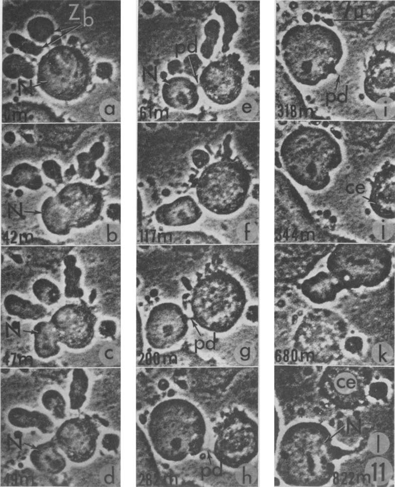
The point was made explicit by time-lapse films of cells held in a thin space under a sheet of cellophane. Carcinoma cells of the KB line blebbed so actively that they seemed to boil, and some of their zeiotic blebs broke up and left droplets of cytoplasm behind; in one cell whose bleb already held the nucleus, constriction rings carried it further out along a lengthening stalk over 66 min (Rose et al., 1963). In three later filmed events the stalk of a bleb parted, and one HeLa bleb that carried the nucleus kept moving by its own blebs for 11 h after its cell apparently died (Rose, 1966). These anecdotal reports, without counts, show that blebs can leave a cell and move on, but not how often (proposed).
Fragments made in the laboratory showed that pieces without a nucleus keep blebbing. Fibroblast microplasts, small anucleate fragments made with the drug cytochalasin B (Carter, 1967), blebbed or ruffled for at least 8 h but never moved from place to place (Albrecht-Buehler, 1980). Of nucleus-free fragments of L929 fibroblasts treated with nocodazole, 70–80% oscillated in shape for at least 8 h, and their bulges emerged through holes that opened in the actin shell under the membrane (Paluch et al., 2005). These fragments tie the blebbing of a fragment to cortex rupture, one of the routes by which blebs start, discussed in Physics of blebs.
Stable blebs that become cytokineplasts
Under confinement between two surfaces a few micrometers apart, many cells extend one large stable bleb that persists and carries the cell forward, the A2 mode of amoeboid migration (Liu et al., 2015). The same authors noted, without showing data, that fragments shed from large blebs moved like whole A2 cells.
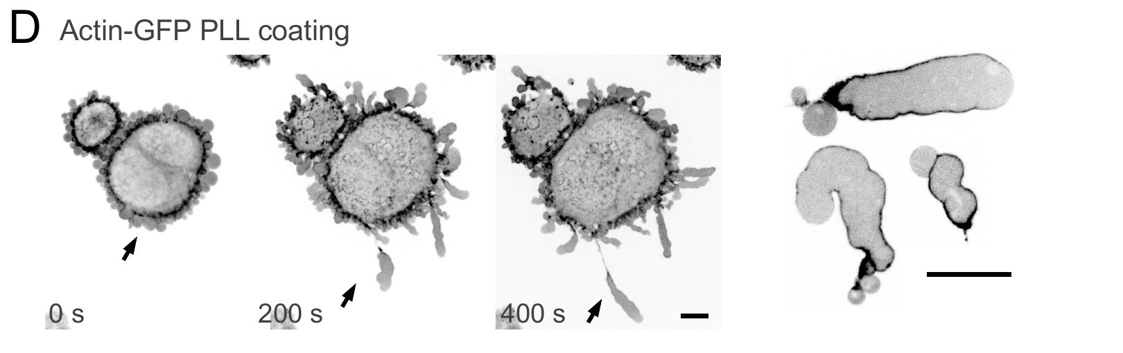
The detachment was then followed in confined HeLa cells. On an adhesive poly-L-lysine coating the cell body stayed in place while stable blebs moved away from it, and the membrane neck at their base narrowed and eventually ruptured. The higher friction on this surface was proposed to let the rearward flow of the bleb’s cortex grip the substrate strongly enough to pull the bleb away (García-Arcos, 2020 ◆). The freed fragments kept the elongated shape and front-to-rear actomyosin gradient of a stable bleb and moved persistently for hours at 5–15 µm/min, and some split again (observations without counts; García-Arcos, 2020 ◆). García-Arcos et al. (2024b ◆) called these fragments cytokineplasts. Their leading edge was almost free of actin filaments and protruded only where cortical actin was sparsest, stalled where actin reached a threshold density and retracted where it was denser still (n = 17 cytoplasts from one replicate), which the authors explain by rigidity percolation (see Physics of blebs). In dense collagen, too, stable blebs moved away from their cell bodies, and the resulting cytokineplasts squeezed through small gaps in the matrix (50 cytoplasts, two replicates). That stable blebs can become motile fragments is strongly supported in HeLa cells, by one laboratory.
These motile fragments bear on an older inference. Because his microplasts blebbed but never translocated, Albrecht-Buehler (1980, p. 6642) proposed that locomotion needs an additional “supervising mechanism”. Fragments made in other ways do migrate: granulocyte fragments released by heating were reported, without data, to migrate toward damaged red cells (Keller and Bessis, 1975), and keratocyte fragments lacking a nucleus, centrioles and most microtubules moved persistently and directionally (Euteneuer and Schliwa, 1984), as do bleb-derived cytokineplasts. A nucleus, centrioles and a full microtubule array are therefore not needed for directed movement, but why the microplasts stayed in place has not been tested in the studies we hold, so the need for a supervising mechanism is unresolved rather than refuted.
Fragments in living tissue
The strongest evidence that cells shed large fragments in an animal comes from the mouse lung. Headley et al. (2016) injected B16 melanoma cells into the circulation and imaged the lung through a window in the chest. Cells that lodged in lung capillaries began within 40 s to shed what the authors called “microscale blebs” and cytoplasts, about 5 µm across, from the tips of cell extensions, and kept doing so for at least 8 h (Headley et al., 2016, p. 513). About four times fewer cells shed fragments in lung slices without flow, the caspase inhibitor Z-VAD did not reduce shedding, and anucleate cytoplasts soon made up most of the tumor material in the lung. Myeloid cells took up the cytoplasts, CD103+ dendritic cells carried tumor material to the lymph node and activated CD8+ T cells, and depleting conventional dendritic cells raised the number of metastases sixfold (Headley et al., 2016).
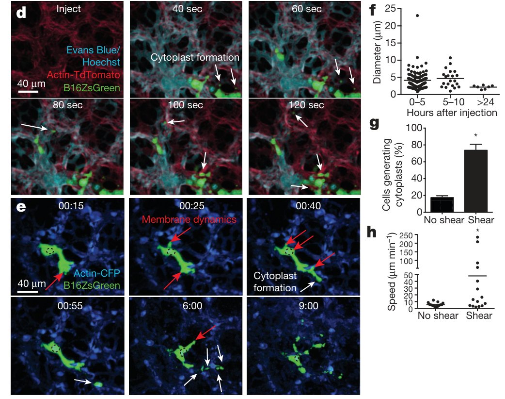
The shedding of these fragments is strongly supported, but their origin from blebs is not: only cytoplasmic and nuclear labels were used, so neither a bleb nor its neck was resolved. Later papers nonetheless cite the lung fragments as bleb-derived. One of them cites a study “in human xenograft tumors” in which cytoplasts “were formed from large blebs” (García-Arcos et al., 2024b ◆, p. 3004), and a review describes fragments “originating from stable blebs” (García-Arcos et al., 2024a ◆, p. 850), whereas Headley et al. filmed injected mouse B16 cells and mention neither xenografts nor stable blebs.
Two other studies in living tissue come closer to the moment of release. In explanted chick embryo skin, a bleb on a melanocyte dendrite pinched off in about 20 s as a vesicle of about 1 µm (one example shown). Of 185 such vesicles, 72.8% lay inside the neighboring keratinocytes, the skin cells that receive pigment, and inhibiting the GTPase Rho reduced both blebbing and release (Tadokoro et al., 2016). In mouse bone marrow imaged in vivo, a free vesicle of about 3.5 µm appeared beside an endothelial bleb 14–23 min after the bleb formed, but with frames 2–3 min apart the separation itself was not resolved (Atkin-Smith et al., 2024).
Apoptotic bodies: one route among several
Apoptotic bodies are the membrane-bound pieces into which dying cells break up and which other cells engulf (Kerr et al., 1972). In some systems, blebs become bodies. An apoptotic neutrophil bleb was filmed detaching, about once per cell every 2 h, and its release needed myosin light-chain kinase but not caspases (Nusbaum et al., 2005). Dying myeloma cells formed a single broad bleb free of organelles, which about half of the cells released (no counts given; Zhang et al., 2005). In Jurkat T cells in which the channel pannexin 1 was blocked, blebs at the tips of thin apoptopodia became bodies of 0.9–5 µm and about 96% of cells fragmented rather than 6%; blocking blebbing abolished the extra bodies, although the separation itself was not shown (Poon et al., 2014).
Other observations separate blebbing from fragmentation. Beaded apoptopodia, strings of beads drawn out from the dying cell, shed bodies of 1–4 µm from THP-1 monocytes and still formed in Jurkat cells whose blebbing was blocked (Atkin-Smith et al., 2015). In pannexin-blocked Jurkat cells, knocking out the kinase ROCK I, which drives apoptotic blebbing, cut the number of apoptotic bodies three- to fivefold, yet blebbing almost vanished in only one of three knockout clones and persisted in about 37–49% of cells of the other two, against about 97% of controls (Tixeira et al., 2020). In our reading, this fits blebs as one source of bodies among several, but it does not show that the bleb is the step that ROCK I controls. Apoptotic cells from zebrafish embryos blebbed in culture, yet none of 115 fragmented, whereas in the embryo the F-actin of resident phagocytes marked all 86 splitting events observed; ROCK inhibition did not block the splits, and mice with a caspase-resistant ROCK I made normal numbers of apoptotic bodies in the thymus (Santavanond et al., 2025). Which route dominates in adult tissues is an open question.
Platelets: buds, proplatelets and blebs
Platelets, the small anucleate fragments that circulate in mammalian blood, are released from megakaryocytes, large polyploid cells of the bone marrow that often lie next to blood vessels (Wright, 1906; Potts et al., 2020). In stained sections, Wright (1906, 1910) saw megakaryocyte buds and slender strands in the vessels that appeared to pinch off into platelets, but on a warm stage he never saw a platelet separate. Whether blebbing takes part in this release has been asked repeatedly since, and the answer depends on what each study meant by a bleb.
The accepted route has long been proplatelet formation, in which a megakaryocyte extends long processes that release platelets from their tips. In mouse bone marrow imaged in vivo, short proplatelets released particles at 1.4 ± 0.3 per minute per megakaryocyte (mean ± s.e.m., 50 cells in 5 mice; Nishimura et al., 2015). Potts et al. (2020) found a different picture. In fixed adult marrow and spleen, about half of the megakaryocytes carried platelet-sized buds and only about 1% formed proplatelets, and in 10–15 min of live imaging, 84% of 45 marrow megakaryocytes budded, losing buds of about 3 µm at about 2 per minute with no proplatelet stage. Budding megakaryocytes in fetal liver showed no active caspase-3, and megakaryocytes lacking the transcription factor NF-E2 largely lost budding while still forming proplatelets.
No bleb has been shown in this budding. Potts et al. (2020) never use the word bleb and did not test pressure, myosin or membrane–cortex attachment; their buds contained F-actin continuous with the actin-rich periphery of the cell, so, in our reading, at least when fixed they were not the cortex-free protrusions that define a growing bleb. Release, moreover, was inferred from buds that vanished between frames. Where the word bleb does appear in this literature, it names something else. In electron micrographs, Italiano et al. (2021) saw megakaryocytes with an organelle-free rim from which protrusions they called ‘blebs’, on shape alone, seemed to pinch off. These held almost no α-granules, the main secretory granules of platelets (fewer than 0.09 per µm², against 2.8 in mature platelets), so the authors read them, and by extension the buds of Potts et al., as microvesicles rather than platelets. In fixed marrow, Ellis et al. (2023) distinguished buds containing the granule protein von Willebrand factor and internal F-actin (2.37 ± 0.43 µm), which they consider platelet precursors, from hollow ‘blebs’ without internal F-actin (3.04 ± 1.39 µm) and from submicron microvesicles. A later review keeps proplatelets as the only route shown to make platelets with all their features (Carminita et al., 2024).
Read together, these studies give no direct evidence that blebbing releases platelets. Bleb routes proposed from electron micrographs in the 1970s (Murata, 1975; Djaldetti et al., 1979) and from megakaryoblastic cell lines (Takeuchi et al., 1998) rest on static images, known to us only from their abstracts. Where megakaryocyte blebbing has been filmed in vivo, it accompanied apoptosis induced with Fas ligand and released no particles (Nishimura et al., 2015). The hollow, actin-free ‘blebs’ of Ellis et al. (2023) look most like growing blebs, but whether megakaryocyte buds or blebs are pressure-driven protrusions where the cortex is absent is an open question.
Large oncosomes: vesicles said to come from blebs
Extracellular vesicles are membrane-bound particles that cells release, from small vesicles of endosomal origin to microvesicles shed from the plasma membrane. Di Vizio et al. (2009) linked a class of large vesicles to blebbing. Epidermal growth factor made DU145 prostate cancer cells bleb within about 5 min, with blebs mostly 0.5–5 µm across, and in time-lapse sequences some blebs retracted while others were extruded and released into the medium. The authors proposed that oncosomes, microvesicles that carry oncogenic signals between cells, may be identical to non-apoptotic blebs.
Di Vizio et al. (2012) then named vesicles of about 1–10 µm large oncosomes. They found them in the plasma of mice carrying human prostate tumor xenografts, in amounts that tracked tumor weight, and in human prostate cancer tissue, more often in metastatic disease. The paper states that the vesicles are released “from pinched membrane blebs” (Di Vizio et al., 2012, p. 1574), but shows the detachment only as one untimed four-frame sequence. Minciacchi et al. (2015) later showed large blebs released as vesicles in one time-lapse movie and found that large oncosomes carry cargo distinct from smaller vesicles. How a bleb would be cut free has been studied mainly in smaller vesicles: in LOX melanoma cells, microvesicles of 300–900 nm were released through a pathway that phosphorylates the myosin light chain, and the myosin inhibitor blebbistatin prevented their formation, although that paper never uses the word bleb (Muralidharan-Chari et al., 2009).
Purified large oncosomes, 4.72 ± 0.28 µm in mean diameter (n = 35), were round and did not bleb, and 54% of them showed no signal with TMRE, a dye that marks mitochondria with a membrane potential (Jeppesen et al., 2025). A perspective nevertheless states that every ectosome, a vesicle shed from the plasma membrane, is a former bleb (Di Vizio et al., 2023). In our reading, the bleb origin of large oncosomes is proposed. Vesicles of bleb size have been filmed leaving blebbing cancer cells a few times, but the fraction of blebs that detach and the events at the neck have not been measured, and in vivo the particles are identified by size and single markers that do not exclude apoptotic bodies or other fragments (Di Vizio et al., 2012).
Blebbisomes: vesicles that bleb without coming from a bleb
Blebbisomes reverse the usual relation between blebs and vesicles. Jeppesen et al. (2025) found exceptionally large vesicles released by cancer cells, with mean diameters from 8.92 ± 0.8 to 14.9 ± 3.0 µm (s.e.m., 167–236 vesicles per line) in three cell lines, that blebbed continuously for at least 72–96 h after release. Blebbisomes have no nucleus but carry mitochondria with a membrane potential similar to that of whole cells, as well as endoplasmic reticulum, Golgi apparatus, endosomes and lysosomes, and they took up smaller vesicles and released their own.
Despite the name, time-lapse imaging showed that blebbisomes “do not originate from a membrane bleb on the cell” (Jeppesen et al., 2025, p. 440). A blebbisome forms instead during a single retraction of the cell: part of the cell stays attached to the substrate, linked to the cell body by a membrane nanotube, and is released when the nanotube is severed. Knocking down non-muscle myosin IIB, a motor that drives retraction, reduced release. Knocking down ESCRT-III proteins, which cut membranes in other budding events, did not, according to the authors, although the figure legend gives 0.001 blebbisomes per cell after CHMP2A knockdown against 0.01 in controls; release was so infrequent that all these comparisons rest on small counts. Blebbing vesicles of this kind were also seen leaving cancer cells in zebrafish embryos and were isolated from normal mouse bone marrow (Jeppesen et al., 2025).
For bleb biology, the point that matters is that a free, anucleate piece of cytoplasm keeps blebbing for days, as nucleus-free fibroblast fragments do for hours (Paluch et al., 2005). How blebbisomes bleb was not studied: no pressure, cortex or membrane–cortex attachment was measured. Formation by retraction rests on one study from one laboratory (proposed), and one commentary already misreports it as “myosin-driven bleb scission” (Gao and Kang, 2025). A vesicle that blebs need not come from a bleb.
Telling fragments apart
Fragment names record how a piece was made or what it does, not where it came from. A cytoplast is any anucleate, membrane-bounded piece of cytoplasm. A cytokineplast is one that keeps its motile machinery, a term coined for heat-released leading edges of neutrophils (Malawista and de Boisfleury Chevance, 1982) and now also used for detached stable blebs (García-Arcos et al., 2024b ◆). A blebbisome is a vesicle that blebs. Released blebs, in turn, are usually classed as microvesicles or large oncosomes by size alone, and sizes overlap across all these classes (Jeppesen et al., 2025).
In our reading, four observations would place a fragment’s origin: live imaging of the separation itself, labels for both membrane and cortex at the neck, the presence or absence of a nucleus and organelles, and behavior after release (proposed). Few studies meet even the first. In our map of the fragmentation literature, 19 of 195 papers image a bleb detaching, and none images the parting of the neck with both membrane and cortex markers (M17). The table summarizes the fragments discussed on this page.
| Fragment | How it forms | Size (system) | Bleb seen leaving the cell? | Sources |
|---|---|---|---|---|
| Merotomy pieces | cut from protists by hand | not recorded here | no | Hofer, 1889 |
| Cytoplasts, microplasts | made with cytochalasin | smallest active microplasts ≈150 µm³ (fibroblasts) | no (made) | Carter, 1967; Albrecht-Buehler, 1980 |
| Heat cytokineplasts | a granule-poor part or the leading edge separates on heating | not recorded here | no | Keller and Bessis, 1975; Malawista and de Boisfleury Chevance, 1982 |
| Hyaline vesicles, blisters, zeiotic blebs | pinched off after alkali damage, torn off by flow, broken into droplets, or the stalk parts | not recorded here | yes, three filmed events (Rose, 1966) | Holtfreter, 1946; Zollinger, 1948; Rose et al., 1963; Rose, 1966 |
| Bleb-derived cytokineplasts | a stable bleb pulls away and its neck ruptures | not recorded here | yes (HeLa; one laboratory) | Liu et al., 2015; García-Arcos, 2020 ◆; García-Arcos et al., 2024b ◆ |
| Lung tumor cytoplasts | shed by tumor cells lodged in capillaries, more with flow | mean 5 µm (mouse lung) | release yes, neck no | Headley et al., 2016 |
| Melanosome vesicles | a dendrite bleb pinches off | ≈1 µm (chick embryo skin) | yes (one event) | Tadokoro et al., 2016 |
| Endothelial vesicles | a free vesicle appears beside a bleb | ≈3.5 µm (mouse marrow) | release not resolved | Atkin-Smith et al., 2024 |
| Apoptotic bodies | blebs at apoptopodia tips, one large bleb, beads, splitting by phagocytes | 0.9–5 µm (Jurkat, PANX1 blocked) | in some systems | Kerr et al., 1972; Zhang et al., 2005; Poon et al., 2014; Santavanond et al., 2025 |
| Platelets | megakaryocyte buds or proplatelets | buds ≈2.4 µm (fixed) to ≈3 µm (live), mouse marrow | no; ‘blebs’ are a separate class | Wright, 1910; Potts et al., 2020; Italiano et al., 2021; Ellis et al., 2023 |
| Large oncosomes | said to be released blebs | 1–10 µm by definition; purified 4.7 µm | a few filmed sequences | Di Vizio et al., 2009; Di Vizio et al., 2012; Jeppesen et al., 2025 |
| Blebbisomes | one retraction; a membrane nanotube is severed | means 8.92–14.9 µm (three cell lines) | no; not from a bleb | Jeppesen et al., 2025 |
Open questions
The first question is what severs the bleb neck. Release has been linked to external flow (Zollinger, 1948; Headley et al., 2016), to actomyosin contraction (Nusbaum et al., 2005; Tadokoro et al., 2016) and, in confined HeLa cells, to the pull of a bleb that propels itself away from an immobilized cell body (García-Arcos, 2020 ◆). None of these has been tied to events at the neck itself, so the mechanism of scission is an open question. The second is how often blebs detach in three-dimensional matrix and in tissue. The only release rate per cell comes from apoptotic neutrophils in culture (Nusbaum et al., 2005), detachment has not been imaged in human tumors, and bleb-derived and lamellipodial cytokineplasts have not been compared.
References (44)
- Albrecht-Buehler G (1980) Autonomous movements of cytoplasmic fragments. Proc Natl Acad Sci USA 77:6639–6643. doi:10.1073/pnas.77.11.6639
- Atkin-Smith GK, Tixeira R, Paone S et al. (2015) A novel mechanism of generating extracellular vesicles during apoptosis via a beads-on-a-string membrane structure. Nat Commun 6:7439. doi:10.1038/ncomms8439
- Atkin-Smith GK, Santavanond JP, Light A et al. (2024) In situ visualization of endothelial cell-derived extracellular vesicle formation in steady state and malignant conditions. Nat Commun 15:8802. doi:10.1038/s41467-024-52867-5
- Carminita E, Becker IC, Italiano JE (2024) What it takes to be a platelet: evolving concepts in platelet production. Circ Res 135:540–549. doi:10.1161/circresaha.124.323579
- Carter SB (1967) Effects of cytochalasins on mammalian cells. Nature 213:261–264. doi:10.1038/213261a0
- Di Vizio D, Kim J, Hager MH et al. (2009) Oncosome formation in prostate cancer: association with a region of frequent chromosomal deletion in metastatic disease. Cancer Res 69:5601–5609. doi:10.1158/0008-5472.can-08-3860
- Di Vizio D, Morello M, Dudley AC et al. (2012) Large oncosomes in human prostate cancer tissues and in the circulation of mice with metastatic disease. Am J Pathol 181:1573–1584. doi:10.1016/j.ajpath.2012.07.030
- Di Vizio D, Schoppet M, Weeraratna A et al. (2023) Blebs and former blebs: from surface protrusions to extracellular vesicles in cancer signalling, anoikis resistance and beyond. J Extracell Biol 2:e112. doi:10.1002/jex2.112
- Djaldetti M, Fishman P, Bessler H et al. (1979) SEM observations on the mechanism of platelet release from megakaryocytes. Thromb Haemost 42:611–620. doi:10.1055/s-0038-1666899 abstract only
- Ellis ML, Alwis I, Smythe R et al. (2023) Megakaryocyte buds are distinct from microvesicles and likely to represent platelet precursors. Blood Adv 7:982–986. doi:10.1182/bloodadvances.2022008850
- Euteneuer U, Schliwa M (1984) Persistent, directional motility of cells and cytoplasmic fragments in the absence of microtubules. Nature 310:58–61. doi:10.1038/310058a0
- Gao Y, Kang T (2025) Blebbisomes as autonomous communication hubs. Extracell Vesicles Circ Nucleic Acids 6:355–359. doi:10.20517/evcna.2025.38
- ◆ García-Arcos JM (2020) Stabilization and motility mechanism of blebs in cancer cells. PhD thesis, Université de Paris (Institut Curie). doi:10.70675/a35575c7z85b5z4ad1z98acz50f8d65ebbac
- ◆ García-Arcos JM, Jha A, Waterman CM et al. (2024a) Blebology: principles of bleb-based migration. Trends Cell Biol 34:838–853. doi:10.1016/j.tcb.2024.02.009
- ◆ García-Arcos JM, Ziegler J, Grigolon S et al. (2024b) Rigidity percolation and active advection synergize in the actomyosin cortex to drive amoeboid cell motility. Dev Cell 59:2990–3007.e7. doi:10.1016/j.devcel.2024.06.023
- Headley MB, Bins A, Nip A et al. (2016) Visualization of immediate immune responses to pioneer metastatic cells in the lung. Nature 531:513–517. doi:10.1038/nature16985
- Hofer B (1889) Experimentelle Untersuchungen über den Einfluss des Kerns auf das Protoplasma. Jena Z Naturwiss 24:105–176.
- Hogue MJ (1919) The effect of hypotonic and hypertonic solutions on fibroblasts of the embryonic chick heart in vitro. J Exp Med 30:617–648. doi:10.1084/jem.30.6.617
- Holtfreter J (1946) Structure, motility and locomotion in isolated embryonic amphibian cells. J Morphol 79:27–62. doi:10.1002/jmor.1050790103
- Italiano JE, Bender M, Merrill-Skoloff G et al. (2021) Microvesicles, but not platelets, bud off from mouse bone marrow megakaryocytes. Blood 138:1998–2001. doi:10.1182/blood.2021012496
- Jeppesen DK, Sanchez ZC, Kelley NM et al. (2025) Blebbisomes are large, organelle-rich extracellular vesicles with cell-like properties. Nat Cell Biol 27:438–448. doi:10.1038/s41556-025-01621-0
- Keller HU, Bessis M (1975) Migration and chemotaxis of anucleate cytoplasmic leukocyte fragments. Nature 258:723–724. doi:10.1038/258723a0
- Kerr JFR, Wyllie AH, Currie AR (1972) Apoptosis: a basic biological phenomenon with wide-ranging implications in tissue kinetics. Br J Cancer 26:239–257. doi:10.1038/bjc.1972.33
- Liu YJ, Le Berre M, Lautenschlaeger F et al. (2015) Confinement and low adhesion induce fast amoeboid migration of slow mesenchymal cells. Cell 160:659–672. doi:10.1016/j.cell.2015.01.007
- Malawista SE, de Boisfleury Chevance A (1982) The cytokineplast: purified, stable, and functional motile machinery from human blood polymorphonuclear leukocytes. J Cell Biol 95:960–973. doi:10.1083/jcb.95.3.960
- Minciacchi VR, You S, Spinelli C et al. (2015) Large oncosomes contain distinct protein cargo and represent a separate functional class of tumor-derived extracellular vesicles. Oncotarget 6:11327–11341. doi:10.18632/oncotarget.3598
- Muralidharan-Chari V, Clancy J, Plou C et al. (2009) ARF6-regulated shedding of tumor cell-derived plasma membrane microvesicles. Curr Biol 19:1875–1885. doi:10.1016/j.cub.2009.09.059
- Murata T (1975) Mechanism of platelet liberation. Tohoku J Exp Med 116:67–75. doi:10.1620/tjem.116.67 abstract only
- Nishimura S, Nagasaki M, Kunishima S et al. (2015) IL-1α induces thrombopoiesis through megakaryocyte rupture in response to acute platelet needs. J Cell Biol 209:453–466. doi:10.1083/jcb.201410052
- Nusbaum P, Lainé C, Bouaouina M et al. (2005) Distinct signaling pathways are involved in leukosialin (CD43) down-regulation, membrane blebbing, and phospholipid scrambling during neutrophil apoptosis. J Biol Chem 280:5843–5853. doi:10.1074/jbc.M413405200
- Paluch E, Piel M, Prost J et al. (2005) Cortical actomyosin breakage triggers shape oscillations in cells and cell fragments. Biophys J 89:724–733. doi:10.1529/biophysj.105.060590
- Poon IKH, Chiu YH, Armstrong AJ et al. (2014) Unexpected link between an antibiotic, pannexin channels and apoptosis. Nature 507:329–334. doi:10.1038/nature13147
- Potts KS, Farley A, Dawson CA et al. (2020) Membrane budding is a major mechanism of in vivo platelet biogenesis. J Exp Med 217:e20191206. doi:10.1084/jem.20191206
- Ranvier L (1890) Des clasmatocytes. C R Hebd Séances Acad Sci 110:165–169.
- Rose GG (1966) Zeiosis. I. Ejection of cell nuclei into zeiotic blebs. J R Microsc Soc 86:87–102. doi:10.1111/j.1365-2818.1966.tb05328.x
- Rose GG, Cattoni M, Pomerat CM (1963) Observations on oral tumors in vitro. 1. Zeiosis versus the nucleus and endoplasmic reticulum in strain KB. J Dent Res 42:38–53. doi:10.1177/00220345630420014401
- Santavanond JP, Atkin-Smith GK, Lozano-González I et al. (2025) Resident phagocytes promote non-cell-autonomous fragmentation of apoptotic cells. Sci Adv 11:eadz5264. doi:10.1126/sciadv.adz5264
- Tadokoro R, Murai H, Sakai KI et al. (2016) Melanosome transfer to keratinocyte in the chicken embryonic skin is mediated by vesicle release associated with Rho-regulated membrane blebbing. Sci Rep 6:38277. doi:10.1038/srep38277
- Takeuchi K, Satoh M, Kuno H et al. (1998) Platelet-like particle formation in the human megakaryoblastic leukaemia cell lines, MEG-01 and MEG-01s. Br J Haematol 100:436–444. doi:10.1046/j.1365-2141.1998.00576.x abstract only
- Tixeira R, Phan TK, Caruso S et al. (2020) ROCK1 but not LIMK1 or PAK2 is a key regulator of apoptotic membrane blebbing and cell disassembly. Cell Death Differ 27:102–116. doi:10.1038/s41418-019-0342-5
- Wright JH (1906) The origin and nature of the blood plates. Boston Med Surg J 154:643–645. doi:10.1056/NEJM190606071542301
- Wright JH (1910) The histogenesis of the blood platelets. J Morphol 21:263–278. doi:10.1002/jmor.1050210204
- Zhang B, Arany Z, Mann D et al. (2005) Partitioning apoptosis: a novel form of the execution phase of apoptosis. Apoptosis 10:219–231. doi:10.1007/s10495-005-6077-4
- Zollinger HU (1948) Cytologic studies with the phase microscope. I. The formation of “blisters” on cells in suspension (potocytosis), with observations on the nature of the cellular membrane. Am J Pathol 24:545–567. PMID 18859358