M041923–1947 · Pressure and gel in the amoeboid cytoplasm, 1923–1982 · established
Amoeboid movement as a pressurized, contracting gel tube
In 1923, without citing each other, Mast and Pantin replaced surface-tension explanations of amoeboid movement with a contracting tube of gelled cytoplasm that pushes the fluid inner cytoplasm forward. The core principle, a contracting outer layer that pressurizes the cytoplasm and pushes it out where the layer gives way, is that of a bleb, and it is now established from measurements in blebbing animal cells (Charras et al., 2005; Tinevez et al., 2009; M12). In amoebae, where the contraction acts and whether a global pressure drives streaming were never settled.
Before
Several observations had undermined the favored explanation, a local fall in surface tension (Mast, 1923). Tracking soot particles, Jennings (1904) found that the surface of an amoeba rolls forward over the front rather than flowing back as on a drop: locomotion was “demonstrably not due to a local decrease in surface tension” (Jennings, 1904, p. 205). An amoeba cutting a paramecium in two exerted far more force than surface tension could supply (Mast and Root, 1916), and in strong cyanide an amoeba’s outer layer dissolved at one spot and the inner cytoplasm burst out there, suggesting a firm elastic layer around a fluid (Hyman, 1917). In our reading, such a burst at a weak spot recalls Ehrenberg’s hernia (Ehrenberg, 1838; M02), but nobody, Mast (1923) complained, had shown how these factors worked together.
What was done
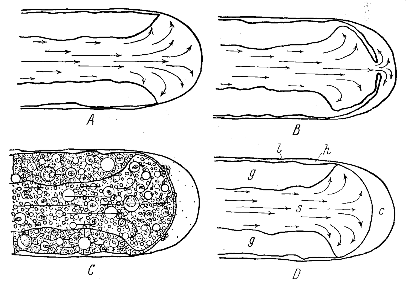
Mast (1923) gave his scheme for Amoeba proteus in four pages without figures or numbers. A fluid core, the plasmasol, lay inside a solid plasmagel under a thin elastic membrane, the plasmalemma, and granules showed the gel forming at the front and dissolving at the rear. The hypertonic sol, he proposed, takes up water and creates an outward pressure, a pseudopod forms where the gel softens, and contraction at the rear drives the sol forward. The pressure was asserted, not measured.
The front was the weak point. At high temperature, Mast reported, the gel at the tip often broke suddenly, the sol rushed through and formed a prominent bulge, and new gel formed beneath it (Mast, 1923). He later likened a starting pseudopod to “a bulge on the surface of an inflated rubber sack” (Mast, 1926, p. 406), and in 1932 he saw “broken edges of a definite layer of gel” where the stream entered the clear tip, or hyaline cap, conceding that the tube was sometimes open at the front (Mast, 1932, p. 322).
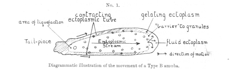
Pantin (1923) reached a similar scheme from three marine amoebae: “a contracting tube of gelated ectoplasm closed at its hind end” (Pantin, 1923, p. 31) that liquefies at the rear, gels again at the front and drives the endoplasm passively forward. The two agreed on the geometry of the motor but not its physics: Mast invoked elastic recoil, Pantin syneresis, the shrinking of a gel as it expels fluid, while admitting that this went beyond his evidence.
Marsland and Brown (1936) softened the gel with high hydrostatic pressure, which made pseudopodia of Amoeba dubia round up within half a second, reversibly, and burst sideways just behind the tip. The gel, they concluded, holds the pseudopod in shape, but its contraction need not drive streaming, since an amoeba capped in oil streamed for hours without changing shape.
| Quantity | Value | System | Source |
|---|---|---|---|
| Force to cut a paramecium | ≈9 mg weight (8.8–9.2); cut 19/30, vs 0/21 at 4.5 mg | Paramecium aurelia, calibrated glass fiber | Mast and Root, 1916 |
| Surface-tension difference needed | ≥383, probably >1118 dyn/cm (protoplasm ≈50) | calculation | Mast and Root, 1916 |
| Speed | 2.06–2.48 µm/s (2 amoebae, 13.4–16.4 °C) | marine limax amoeba | Pantin, 1923 |
| Streaming stops; pseudopodia round up | ≈250 atm; ≈400 atm, sphere in 0.38 s (1 cell); n not stated | A. dubia, rapid compression | Marsland and Brown, 1936 |
| Lateral wall ruptures | 4–6 per min at 100–204 atm; n not stated | Amoeba held at pressure | Marsland and Brown, 1936 |
What it changed
By 1947 the gel tube had displaced surface tension: reversible sol–gel changes were established, and most investigators attributed movement to contraction of the plasmagel (De Bruyn, 1947). Mast’s scheme framed the first measurements of pressure and viscosity in moving amoebae (M05), and the gel was later recast as a network of actin filaments (M06).
Bleb research returned to these ideas. García-Arcos et al. (2024b ◆), citing Hyman, Pantin and Mast, described the actin transition in stable blebs as rigidity percolation, the point at which a network becomes connected enough to transmit force. In our reading, Mast’s breaking tip follows the sequence of a bleb started by cortex rupture, with the gel breaking, the membrane bulging and new gel forming beneath (proposed), as may Hyman’s cyanide burst and Marsland and Brown’s lateral ruptures, although none of these authors used the word bleb.
Later work and open questions
The scheme was disputed from the start: Jennings (1904) had ruled out a general pressure from behind, since amoebae are thickest at the rear, and De Bruyn (1947) rejected Mast’s osmotic recoil because of that open front. Later measurements also questioned pressure as the driver (Allen and Roslansky, 1959; Yanai et al., 1996; M05).
Whether Mast’s breaking tip is a bleb has not been tested. The only time-resolved record we know of shows the front cortical layer of A. proteus detaching from the membrane about every 2 s (2.17 ± 1.22 s, mean ± s.d., 304 cycles) rather than rupturing (Grębecki, 1990), although rupture can start blebs in other systems (M15). Both 1923 schemes rest on qualitative observations of a few species, and Mast’s heat-induced rupture may have been injury.
Two questions remain open in amoebae: whether the pressure that drives the flow is global, as Mast assumed, or local (M12), and what weakens the front, whether softening of the gel, detachment of the membrane or a network falling below a connectivity threshold (open question).
References (16)
- Allen RD, Roslansky JD (1959) The consistency of ameba cytoplasm and its bearing on the mechanism of ameboid movement. I. An analysis of endoplasmic velocity profiles of Chaos chaos (L.). J Biophys Biochem Cytol 6:437–446. doi:10.1083/jcb.6.3.437
- Charras GT, Yarrow JC, Horton MA et al. (2005) Non-equilibration of hydrostatic pressure in blebbing cells. Nature 435:365–369. doi:10.1038/nature03550
- De Bruyn PPH (1947) Theories of amoeboid movement. Q Rev Biol 22:1–24. doi:10.1086/395577
- Ehrenberg CG (1838) Die Infusionsthierchen als vollkommene Organismen. Leipzig: Voss. pp. 125–129. No DOI.
- ◆ García-Arcos JM, Ziegler J, Grigolon S et al. (2024b) Rigidity percolation and active advection synergize in the actomyosin cortex to drive amoeboid cell motility. Dev Cell 59:2990–3007.e7. doi:10.1016/j.devcel.2024.06.023
- Grębecki A (1990) Dynamics of the contractile system in the pseudopodial tips of normally locomoting amoebae, demonstrated in vivo by video-enhancement. Protoplasma 154:98–111. doi:10.1007/BF01539837
- Hyman LH (1917) Metabolic gradients in amoeba and their relation to the mechanism of amoeboid movement. J Exp Zool 24:55–99. doi:10.1002/jez.1400240104
- Jennings HS (1904) Contributions to the study of the behavior of lower organisms. Carnegie Institution of Washington Publication 16, Washington. doi:10.5962/bhl.title.30720
- Marsland DA, Brown DES (1936) Amoeboid movement at high hydrostatic pressure. J Cell Comp Physiol 8:167–178. doi:10.1002/jcp.1030080205
- Mast SO (1932) Locomotion in Amoeba proteus (Leidy). Protoplasma 14:321–330 (received 9 May 1931). doi:10.1007/BF01604911
- Mast SO (1923) Mechanics of locomotion in Amoeba. Proc Natl Acad Sci USA 9:258–261. doi:10.1073/pnas.9.7.258
- Mast SO, Root FM (1916) Observations on ameba feeding on rotifers, nematodes and ciliates, and their bearing on the surface-tension theory. J Exp Zool 21:33–49. doi:10.1002/jez.1400210103
- Mast SO (1926) Structure, movement, locomotion, and stimulation in amoeba. J Morphol 41:347–425. doi:10.1002/jmor.1050410205
- Pantin CFA (1923) On the physiology of amoeboid movement. I. J Mar Biol Assoc UK 13:24–69. doi:10.1017/S0025315400010924
- Tinevez JY, Schulze U, Salbreux G et al. (2009) Role of cortical tension in bleb growth. Proc Natl Acad Sci USA 106:18581–18586. doi:10.1073/pnas.0903353106
- Yanai M, Kenyon CM, Butler JP et al. (1996) Intracellular pressure is a motive force for cell motion in Amoeba proteus. Cell Motil Cytoskeleton 33:22–29. doi:10.1002/(SICI)1097-0169(1996)33:1<22::AID-CM3>3.0.CO;2-K