M202015 · Blebs in migration · established
Confinement switches many cells to stable-bleb migration
In 2015, four studies showed that confinement, combined with low adhesion and high contractility, turns slow cells of many types into fast amoeboid cells (Liu et al., 2015; Ruprecht et al., 2015). The fastest are led by one large, persistent bleb, called a stable bleb or leader bleb (Logue et al., 2015), and are propelled by a rearward flow of the cortex that grips the walls by non-specific friction (Bergert et al., 2015). The switch is established in vitro, and the friction coupling is strongly supported.
Before
Charras and Paluch (2008) had proposed that confined blebbing cells could push on the walls without specific adhesion (M19). Rounded breast cancer cells crossing Matrigel then pointed to a mechanism. Their cortex flowed rearward toward a contractile rear, carrying matrix beads with it, and migration was 45–65% slower without β1 integrin; blebs appeared only at the rear, which the authors judged a by-product (Poincloux et al., 2011). A model showed how friction could turn such flow into movement of a round cell (Hawkins et al., 2011), and a non-adherent, blebbing subline of Walker carcinosarcoma cells migrated only when confined (Bergert et al., 2012). Still missing were a way to induce the mode across cell types, a direct test of friction and a molecular regulator.
What was done
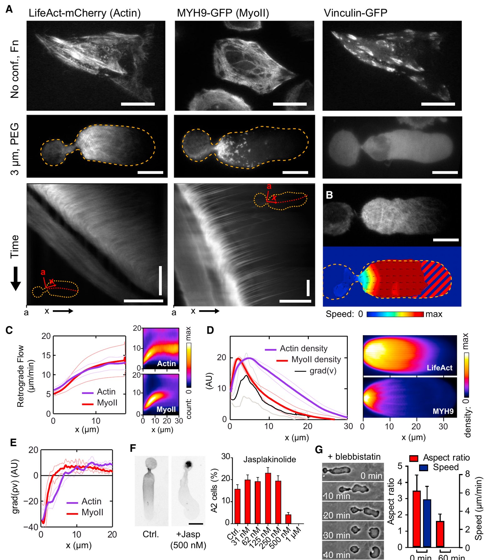
Liu et al. (2015) confined cells under a roof, mostly 3 or 5 µm high, on non-adhesive polyethylene glycol (PEG). At 3 µm, fibroblasts and 20 other cell types, all but normal epithelial cells, produced a fast sub-population, about 10–40% of cells per line, in two modes, A1 and A2. A2 cells, elongated with a trailing uropod, needed focal adhesions to be absent and disappeared with blebbistatin. They entered this mode through a large bleb that took in most of the cytoplasm and the nucleus, and whose cortex, absent at the front and dense at the rear, flowed rearward.
Ruprecht et al. (2015) reached the same mode by raising contractility. Serum or lysophosphatidic acid turned zebrafish progenitor cells into stable-bleb cells, a polarization that needed myosin II and Rho-associated kinase (ROCK) and began when cortical flow away from a bleb prevented its retraction. In embryos, cells with active RhoA, or cells transplanted near wounds, became stable-bleb-like.
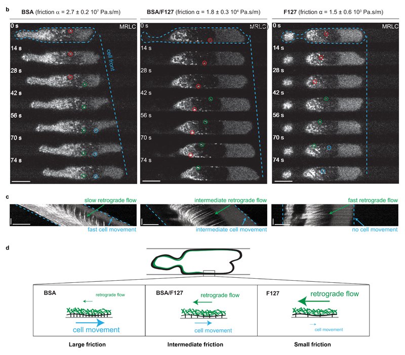
In a simple picture, the cortex of such cells behaves like a conveyor belt: it assembles at the front of the bleb, flows rearward and is taken apart at the rear, where myosin pulls on it. If the belt grips the walls, the cell moves; if it slips, the belt runs in place. Bergert et al. (2015) tested this with Walker cells in channels whose coatings set the friction. On a high-friction coating the cortex stayed nearly fixed relative to the wall and the cell moved; on the lowest-friction coating the cortex flowed but the cell stood still. The authors concluded that “actin cortex flows drive cell movement via non-specific substrate friction” (p. 524), with stresses, estimated from friction on unpolarized cells, of about 1 Pa or less.
Logue et al. (2015) added a molecular regulator. Confined melanoma cells formed one elongated leader bleb beyond a thin neck, and both the size of this bleb and the cells’ migration depended on Eps8, a protein that bundles and caps actin filaments.
| Quantity | Value | System | Source |
|---|---|---|---|
| Speed, A2 / A1 / mesenchymal | 5.3 ± 1.5 / 1.7 ± 0.4 / 0.234 ± 0.09 µm/min (s.e.m.; 2–46 cells per line and mode) | cell lines, 3 µm, PEG | Liu et al., 2015, Fig. 4C |
| Rearward cortical flow, A2 | 15.8 ± 3.7 µm/min (s.d., n = 11), cell frame | HeLa, 3 µm, PEG | Liu et al., 2015, Results |
| Stable-bleb cell speed | 17.1 ± 0.6 µm/min (s.e.m., n = 67); in vivo 8.5 ± 0.3 µm/min (s.e.m., n = 15) | zebrafish progenitors; transplanted into embryos | Ruprecht et al., 2015, Figs 5E–F, 7F–G |
| Friction coefficient; cell speed | bovine serum albumin (BSA) 2.7 ± 0.2 × 10⁷ Pa·s/m, 7.6 ± 0.5 µm/min (n = 18); Pluronic F127 1.5 ± 0.6 × 10³ Pa·s/m, no movement (s.e.m.) | Walker cells in channels | Bergert et al., 2015, Fig. 2b |
| Cortical tension; pressure, control vs Eps8 knockdown | 1,594 ± 80 vs 1,207 ± 42 pN/µm; 428 ± 24 vs 339 ± 13 Pa (s.e.m.; n = 13, 20) | rounded A375 melanoma cells, atomic force microscopy | Logue et al., 2015, Fig. 3C–D |
What it changed
The 2015 work made stable-bleb migration a behavior that many cells can adopt, and it raised the question of how cells sense confinement. In zebrafish progenitors the stable-bleb fraction rose from about 23% at 13 µm to about 58% at 7 µm of confinement, and deformation of the nucleus was proposed as the sensor (Venturini et al., 2020); a companion study in HeLa cells found the same route (Lomakin et al., 2020 ◆).
Later work and open questions
Stable blebs of confined HeLa cells have since been followed at the level of single actin filaments: the front lay below the connectivity (percolation) threshold, the middle moved as a connected network, and myosin IIA was confined to the rear (García-Arcos et al., 2024b ◆; see Physics of blebs).
Several findings qualify the 2015 picture. Adhesion is not always absent: stable-bleb progenitors also moved on fibronectin with adhesion foci (Ruprecht et al., 2015), and rounded melanoma cells in dense collagen formed adhesions between blebs (Driscoll et al., 2024 ◆). In unperturbed zebrafish larvae, T cells moving in a 3 µm gap between layers of keratinocytes kept actin at the rear and pushed a bulge of cytoplasm ahead; in skin explants their actin flowed rearward relative to the cell, and myosin II or ROCK inhibitors slowed them (Robertson et al., 2025). Their bleb identity, however, was inferred without membrane or cortex markers, so the mode in unperturbed tissue remains a proposal.
How cells sense confinement seems to differ between cells and tissues. Neutrophils in narrow channels kept their speed without the nuclear route, although blebs were not examined (Deygas et al., 2026), and chick germ cells needed it in one tissue only (Morimoto et al., 2026 preprint). Piezo1 is implicated in confined melanoma cells (Kuang et al., 2025 preprint), yet activating Piezo1 stopped induced blebbing in breast cancer cells (O’Callaghan et al., 2022). Whether a common sensor exists is an open question, as is how force is transmitted in tissue. See also Blebs in cell migration.
References (17)
- Bergert M, Chandradoss SD, Desai RA et al. (2012) Cell mechanics control rapid transitions between blebs and lamellipodia during migration. Proc Natl Acad Sci USA 109:14434–14439. doi:10.1073/pnas.1207968109
- Bergert M, Erzberger A, Desai RA et al. (2015) Force transmission during adhesion-independent migration. Nat Cell Biol 17:524–529. doi:10.1038/ncb3134
- Charras G, Paluch E (2008) Blebs lead the way: how to migrate without lamellipodia. Nat Rev Mol Cell Biol 9:730–736. doi:10.1038/nrm2453
- Deygas M, Bernard M, Nivoit P et al. (2026) Adaptive motility enables neutrophils to rapidly navigate confined capillaries. Sci Immunol 11:eaea1426. doi:10.1126/sciimmunol.aea1426
- ◆ Driscoll MK, Welf ES, Weems A et al. (2024) Proteolysis-free amoeboid migration of melanoma cells through crowded environments via bleb-driven worrying. Dev Cell 59:2414–2428.e8. doi:10.1016/j.devcel.2024.05.024
- ◆ García-Arcos JM, Ziegler J, Grigolon S et al. (2024b) Rigidity percolation and active advection synergize in the actomyosin cortex to drive amoeboid cell motility. Dev Cell 59:2990–3007.e7. doi:10.1016/j.devcel.2024.06.023
- Hawkins RJ, Poincloux R, Bénichou O et al. (2011) Spontaneous contractility-mediated cortical flow generates cell migration in three-dimensional environments. Biophys J 101:1041–1045. doi:10.1016/j.bpj.2011.07.038
- Kuang S, Abrenica A, Kar N et al. (2025) Targeting cholesterol-dependent Piezo1 activation impairs amoeboid migration in melanoma cells. bioRxiv. doi:10.1101/2025.07.11.664494 preprint (PNAS 2026 version: doi:10.1073/pnas.2607369123, abstract only)
- Liu YJ, Le Berre M, Lautenschlaeger F et al. (2015) Confinement and low adhesion induce fast amoeboid migration of slow mesenchymal cells. Cell 160:659–672. doi:10.1016/j.cell.2015.01.007
- Logue JS, Cartagena-Rivera AX, Baird MA et al. (2015) Erk regulation of actin capping and bundling by Eps8 promotes cortex tension and leader bleb-based migration. eLife 4:e08314. doi:10.7554/eLife.08314
- ◆ Lomakin AJ, Cattin CJ, Cuvelier D et al. (2020) The nucleus acts as a ruler tailoring cell responses to spatial constraints. Science 370:eaba2894. doi:10.1126/science.aba2894
- Morimoto M, Kamei Y, Morita M et al. (2026) Environment-specific mechanosensing preserves a common bleb-based migratory program across diverse embryonic environments. bioRxiv. doi:10.64898/2026.08.11.744326 preprint
- O’Callaghan P, Engberg A, Eriksson O et al. (2022) Piezo1 activation attenuates thrombin-induced blebbing in breast cancer cells. J Cell Sci 135:jcs258809. doi:10.1242/jcs.258809 (earlier version: bioRxiv 2020, doi:10.1101/2020.04.30.068338 preprint, superseded)
- Poincloux R, Collin O, Lizárraga F et al. (2011) Contractility of the cell rear drives invasion of breast tumor cells in 3D Matrigel. Proc Natl Acad Sci USA 108:1943–1948. doi:10.1073/pnas.1010396108
- Robertson TF, Schrope J, Zwick Z et al. (2025) Live imaging in zebrafish reveals tissue-specific strategies for amoeboid migration. Development 152:dev204351. doi:10.1242/dev.204351
- Ruprecht V, Wieser S, Callan-Jones A et al. (2015) Cortical contractility triggers a stochastic switch to fast amoeboid cell motility. Cell 160:673–685. doi:10.1016/j.cell.2015.01.008
- Venturini V, Pezzano F, Català Castro F et al. (2020) The nucleus measures shape changes for cellular proprioception to control dynamic cell behavior. Science 370:eaba2644. doi:10.1126/science.aba2644