M101995–1999 · The regulated bleb, 1995–2003 · established
A bleb is membrane freed from the cortex
By the early 1990s it was unclear whether a bleb contains actin and what its membrane is attached to. Work in 1995–1999 gave the answer that still defines a bleb: the plasma membrane moves away from the cortex, the actomyosin network beneath it, which stays behind, and the bleb expands before new actin assembles under its membrane and stops growing as that cortex forms (established). Their loosely attached membrane also made blebs probes of membrane tension and intracellular pressure, with values that depend strongly on method.
Before
Membrane separation had been seen early but read differently: Zollinger (1948) took the clear contents of blisters that detached the membrane of suspended cells for cortex dissolved to a fluid sol. Indirect evidence suggested that bleb membrane lacks cytoskeletal support. Membrane receptors diffused about 100 times faster on induced blebs than on the intact surface (Tank et al., 1982), and fibroblast blebs lacked F-actin staining (Barak and Webb, 1982). Neutrophil blebs grew at a mean 2.4 µm/s (14 blebs), too fast, Keller and Niggli (1993) judged, for growing actin filaments to push them, but their fixed blebs, some lined with F-actin and some almost bare, could not show the order of events.
What was done
Melanoma cells lacking actin-binding protein (ABP), the actin cross-linker filamin, blebbed over their whole surface, and restoring ABP (in the subline A7) confined blebbing to transient local blebs (Cunningham et al., 1992). Cunningham (1995) filmed the blebs of the deficient M2 line: each grew at a constant volumetric rate for 4–7 s and stopped abruptly, at a final volume of 5–125 µm³ proportional to that rate (about 20 blebs, without statistics). Injected phalloidin, which binds actin filaments, stained stable but not expanding blebs, and Cunningham concluded that “a polymerized actin structure is not present initially” (p. 1589) but appears later and stops expansion.
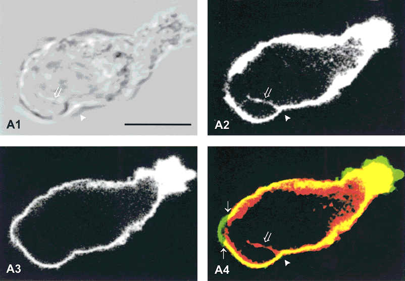
Keller and Eggli (1998) asked where the cortex goes. In fixed, colchicine-treated Walker 256 carcinosarcoma cells, a tumor line that moves by blebbing, the membrane bulged where the cortex was thin and fenestrated, “whereas the actin layer stays in place” (p. 183), and elsewhere it had left a largely intact actin layer behind (figure). The authors read this as forward movement of membrane rather than retraction of actin, in a cycle of detachment, new actin assembly on the membrane and disassembly of the old layer.
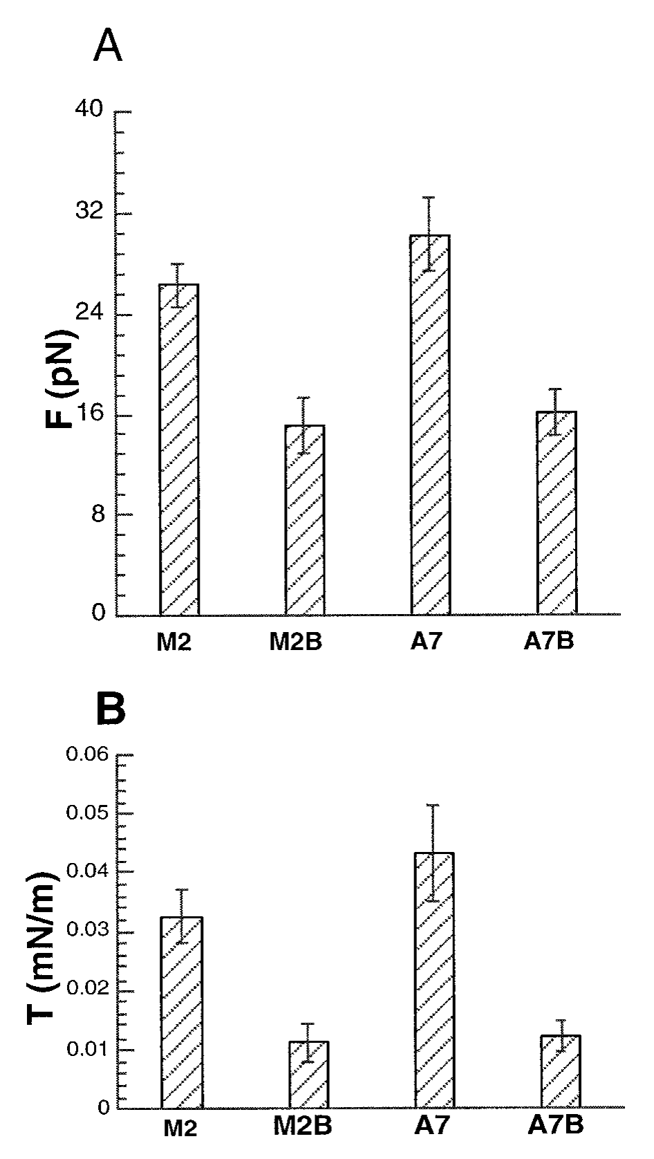
Dai and Sheetz (1999) used laser tweezers to pull thin membrane tubes, or tethers, from blebs and from cortex-supported membrane of melanoma cells. The force needed to hold a tether reflects the membrane’s tension plus its attachment to the cortex, and tethers from blebs needed about half the force. Assuming that a bleb tether reports in-plane tension alone, the authors obtained 11–12 pN/µm for bleb membrane and assigned the rest of the apparent tension of supported membrane to membrane–cortex attachment. Laplace’s law, relating tension and curvature to pressure, then gave an intracellular pressure of about 20 Pa (table), a single-study value (proposed).
| Quantity | Value | System | Source |
|---|---|---|---|
| Applied suction that started blebbing | 1.7 ± 0.7 cm H₂O (range 0.9–3, ≈90–290 Pa by our conversion; mean ± s.d.; 10 of 15 cells blebbed) at 37 °C; 2.8 ± 0.3 cm H₂O (2 of 15) at room temperature | Walker cells, 5–8 µm micropipettes | Rentsch and Keller, 2000, Results |
| Tether force and apparent tension, supported vs bleb membrane | M2 ≈26 vs ≈15 pN (n = 40, 94), ≈32 vs 11 pN/µm; A7 ≈30 vs ≈16 pN (n = 26 each), ≈43 vs 12 pN/µm; from plot, error type not stated | M2 and A7 melanoma cells, laser-tweezer tethers | Dai and Sheetz, 1999, Fig. 5 |
| Intracellular pressure (Laplace’s law) | 20.7 ± 5.9 Pa (M2), 24.2 ± 8.0 Pa (A7); s.d., n not stated | melanoma cells, ~6 h after plating; bleb tension and radius | Dai and Sheetz, 1999, Results |
What it changed
Separation from the cortex became the working definition of a bleb, and later work confirmed the sequence in live cells. Tagged actin was faint on the rims of extending neuroblastoma blebs and brightened as retraction began, although dilution on an expanding surface could hide early actin (Hagmann et al., 1999). Suction alone raised blebs on Walker cells that left actin layers behind (Rentsch and Keller, 2000), and in untreated Walker cells an old actin layer stayed at the base of 88.7 ± 7.6% of blebs (mean ± s.d., 3–4 experiments; Keller et al., 2002). Charras (2008) condensed this into a functional definition: a bleb is membrane transiently decoupled from the cortex, with nucleation requiring actomyosin contraction.
Later work and open questions
How membrane and cortex first separate depends on the system (rupture versus detachment). Untreated Walker cells mostly fit detachment, but under 0.1 µM latrunculin A, which depolymerizes actin, 23.5 ± 10.5% of their blebs (mean ± s.d.) formed where the cortex had first disassembled (Keller et al., 2002). In nocodazole-treated fragments of L929 fibroblasts a hole opened in the actin shell before an actin-free bulge grew, and in whole cells local cytochalasin D or latrunculin A raised a bulge at the treated site (8 of 12 and 7 of 11 cells) but not after the myosin inhibitor blebbistatin (0 of 9). The authors read this as contraction-driven rupture of the cortex, although their bulge grew to cell size (Paluch et al., 2005; M15).
Bleb membrane tension is less settled. Dai and Sheetz themselves called their value an upper limit, and in the same M2 cells thermal fluctuations of bleb membrane gave 0.8–1.0 pN/µm, about ten times less (Charras et al., 2008). In our reading the two methods probe different things, a pulled tube against the undulations of an intact surface, and which gives the in-plane tension of a bleb is an open question. The pressure estimate rests on a relation between bleb size and tether force tested on four blebs of one cell, larger than those measured, and from these points we calculate about 40–45 Pa, twice the reported mean. Whether one bleb reports the pressure of the whole cell is taken up on M12.
References (14)
- Barak LS, Webb WW (1982) Diffusion of low density lipoprotein-receptor complex on human fibroblasts. J Cell Biol 95:846–852. doi:10.1083/jcb.95.3.846
- Charras GT (2008) A short history of blebbing. J Microsc 231:466–478. doi:10.1111/j.1365-2818.2008.02059.x
- Charras GT, Coughlin M, Mitchison TJ et al. (2008) Life and times of a cellular bleb. Biophys J 94:1836–1853. doi:10.1529/biophysj.107.113605
- Cunningham CC (1995) Actin polymerization and intracellular solvent flow in cell surface blebbing. J Cell Biol 129:1589–1599. doi:10.1083/jcb.129.6.1589
- Cunningham CC, Gorlin JB, Kwiatkowski DJ et al. (1992) Actin-binding protein requirement for cortical stability and efficient locomotion. Science 255:325–327. doi:10.1126/science.1549777
- Dai J, Sheetz MP (1999) Membrane tether formation from blebbing cells. Biophys J 77:3363–3370. doi:10.1016/S0006-3495(99)77168-7
- Hagmann J, Burger MM, Dagan D (1999) Regulation of plasma membrane blebbing by the cytoskeleton. J Cell Biochem 73:488–499. doi:10.1002/(SICI)1097-4644(19990615)73:4<488::AID-JCB7>3.0.CO;2-P
- Keller H, Eggli P (1998) Protrusive activity, cytoplasmic compartmentalization, and restriction rings in locomoting blebbing Walker carcinosarcoma cells are related to detachment of cortical actin from the plasma membrane. Cell Motil Cytoskeleton 41:181–193. doi:10.1002/(SICI)1097-0169(1998)41:2<181::AID-CM8>3.0.CO;2-H
- Keller H, Rentsch P, Hagmann J (2002) Differences in cortical actin structure and dynamics document that different types of blebs are formed by distinct mechanisms. Exp Cell Res 277:161–172. doi:10.1006/excr.2002.5552
- Keller HU, Niggli V (1993) Colchicine-induced stimulation of PMN motility related to cytoskeletal changes in actin, α-actinin, and myosin. Cell Motil Cytoskeleton 25:10–18. doi:10.1002/cm.970250103
- Paluch E, Piel M, Prost J et al. (2005) Cortical actomyosin breakage triggers shape oscillations in cells and cell fragments. Biophys J 89:724–733. doi:10.1529/biophysj.105.060590
- Rentsch PS, Keller H (2000) Suction pressure can induce uncoupling of the plasma membrane from cortical actin. Eur J Cell Biol 79:975–981. doi:10.1078/0171-9335-00124
- Tank DW, Wu ES, Webb WW (1982) Enhanced molecular diffusibility in muscle membrane blebs: release of lateral constraints. J Cell Biol 92:207–212. doi:10.1083/jcb.92.1.207
- Zollinger HU (1948) Cytologic studies with the phase microscope. I. The formation of “blisters” on cells in suspension (potocytosis), with observations on the nature of the cellular membrane. Am J Pathol 24:545–567. PMID 18859358