Blebs in dying and injured cells
What drives blebbing in apoptotic, necrotic and injured cells, how these blebs differ from those of living cells, whether they help the cell, and when blebbing merely marks death.
Blebbing has long been read as a sign of a cell in trouble. The earliest definition of the word bleb that we found describes dying cells: Hogue (1919) applied it to clear, rounded bulges of chick fibroblasts dying in concentrated saline (M03). Today blebbing is best known as a feature of apoptosis, the programmed cell death carried out by proteases called caspases, but it also appears in cells injured by lack of oxygen, toxins or heat. This page asks what drives these blebs, whether they help or merely mark the dying cell, and how they become apoptotic bodies.
From zeiosis to apoptosis
In the 1950s and 1960s, the blebbing of cultured cells was often called zeiosis, ‘boiling over’, a term Costero and Pomerat (1951) coined for stalked blebs on the dendrites of cultured neurons. Zeiosis was usually reversible, although some cells died in it (Rose et al., 1963; Rose, 1966). One early explanation was that a fall in ATP relaxed the cell surface and let it bulge (Lettré’s hypothesis, as summarized by Dornfeld and Owczarzak, 1958). Cells treated with cytochalasin D pointed the other way: they formed knobs of cytoplasm as they contracted (Miranda et al., 1974a), and energy poisons cut this zeiosis from 70% to 2% of HeLa cells (Godman et al., 1975). These results indicate that drug-induced blebbing, at least, is an active, energy-consuming result of cortical contraction (M08).
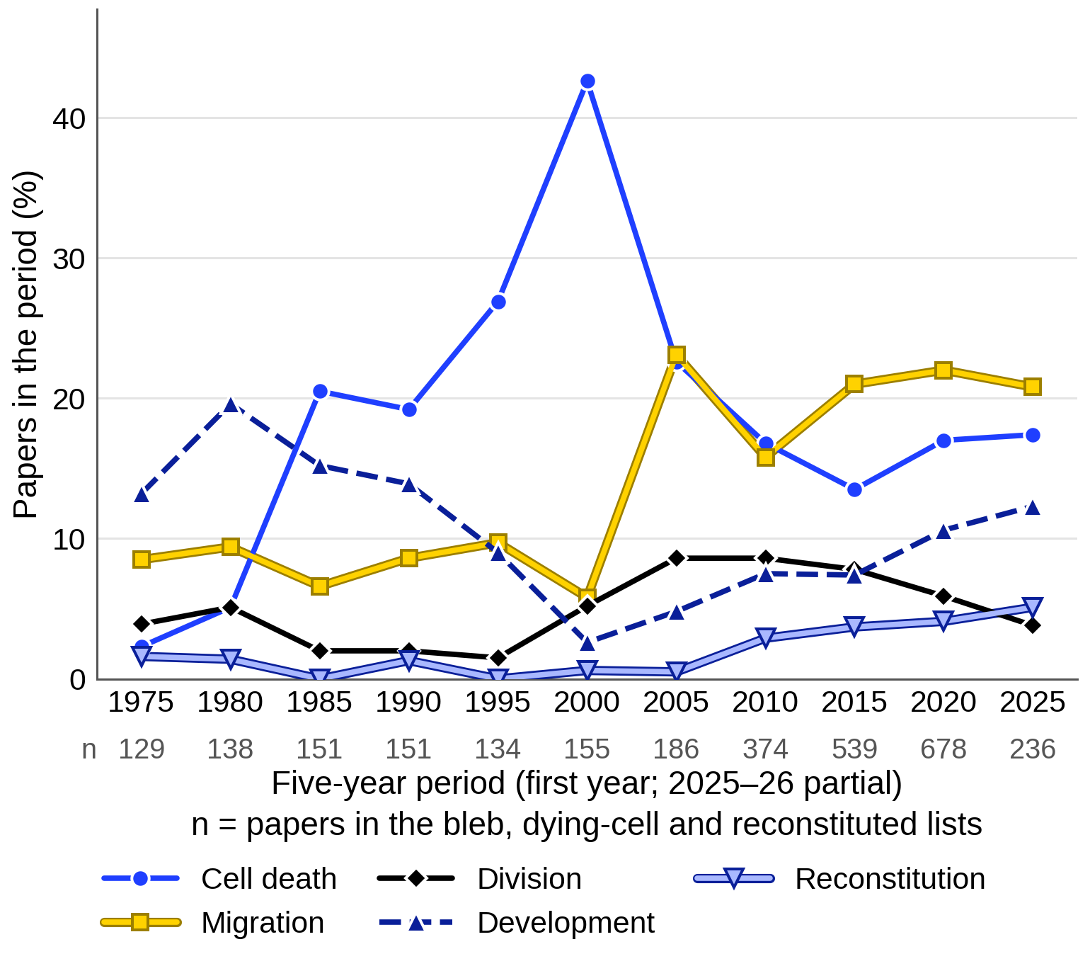
The term passed into the study of injury (Lemasters et al., 1983), and cell death became a major setting of bleb research. It was the setting of 43% of the bleb papers we counted for 2000–2004, its peak, and since 2015 it has been the second most common after migration.
The contractile engine of apoptotic blebbing
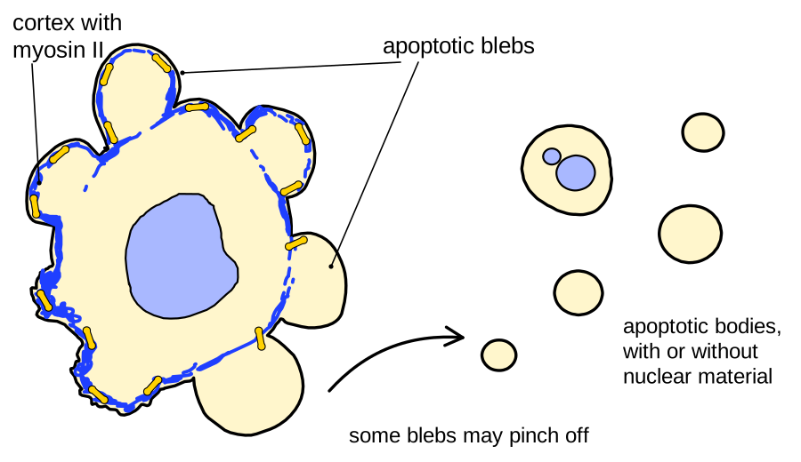
Apoptotic blebbing is driven by the same machinery as the blebs of living cells. Myosin II motors contract the cortex, the network of actin filaments under the membrane; the resulting tension pressurizes the cytoplasm, and the pressure inflates the membrane wherever it is not held back (Tinevez et al., 2009; see Physics of blebs). In dying cells the evidence concerns the contractile step; the pressure has not been measured in the apoptosis studies we hold.
The first clue came from serum-deprived PC12 cells kept alive with caspase inhibitors, which went on blebbing for hours to days. Only inhibitors of myosin light-chain kinase (MLCK), which activates myosin II, reduced the blebbing, and the myosin light chain was 3–5-fold more phosphorylated in blebbing cultures (Mills et al., 1998). Caspases supply the activation by cleaving the Rho-associated kinase ROCK I (Coleman et al., 2001; Sebbagh et al., 2001). The truncated kinase was 7.8 ± 0.5-fold more active (s.e.m., 5 experiments) and made cells bleb even when caspases were blocked (Coleman et al., 2001). Knocking out ROCK I almost abolished apoptotic blebbing in one of three Jurkat clones and reduced it in the other two (Tixeira et al., 2020). Because the 1998 evidence rested on inhibitors with overlapping targets, it is the truncated kinase and the knockouts that make the dependence of apoptotic blebbing on actomyosin contraction strongly supported (M11).
The same activation drives stress blebbing from which cells recover, as in pancreatic acini over-stimulated with the hormone cholecystokinin (Torgerson and McNiven, 1998).
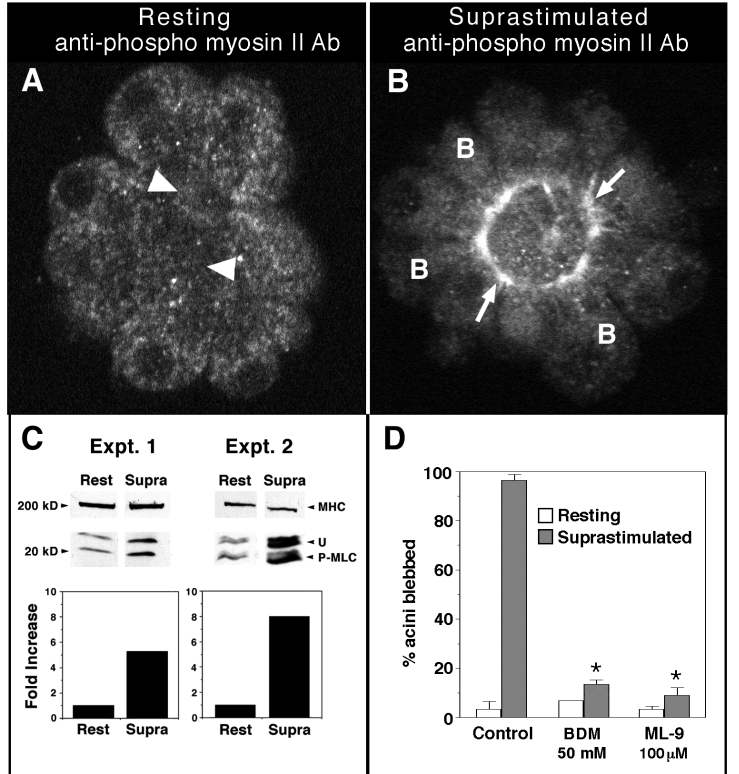
Upstream of this shared core, the signals differ. Blebbing needed Rho in caspase-inhibited PC12 cells (Mills et al., 1998) and the kinase p38 rather than caspases in cisplatin-treated Rat-1 cells (Deschesnes et al., 2001), but caspases and not Rho in other systems (Jänicke et al., 1998; Coleman et al., 2001; Sebbagh et al., 2001). These studies differ in cell type, stimulus and whether caspases were blocked, and Sebbagh et al. (2001) proposed that adherent cells may bleb through Rho as they round up. Even the role of myosin activity varies: in endothelial cells treated with combretastatin A-4-phosphate, MLCK and myosin ATPase inhibitors raised blebbing from 16 ± 4.5% (s.d., 4 experiments) to about 39–44% of cells (Kanthou and Tozer, 2002).
How apoptotic blebs differ from blebs of living cells
The blebs themselves change as apoptosis proceeds. In DLD1 colon cancer cells, the GTPase Rnd3 marked expanding blebs and active RhoA retracting ones in both migrating and apoptotic cells. Apoptotic blebs, however, grew with time, from about 5 µm² early in apoptosis to 45 µm² late, against 14 µm² in migrating cells (10 blebs each, read from plot), and late blebs retracted slowly. Because active ROCK I made the blebs of migrating cells smaller and an active lipid scramblase, which mixes the lipids of the two membrane leaflets, made them larger, the authors propose that ROCK I produces the small early blebs and lipid scrambling the large late ones (Aoki et al., 2020; one cell line).
Fixed images are less informative. Apoptotic blebs were almost free of organelles and of F-actin except at their base (Laster and Mackenzie, 1996), and lacked membrane-bound ERM proteins and the hyaluronan coat of the cell surface, but so did blebs raised by contraction in healthy cells (Le et al., 2024), and expanding blebs of living cells also lack polymerized actin at first (Cunningham, 1995). In our reading, no criterion visible in a single image yet separates apoptotic blebs from the dynamic blebs of living cells (open question).
Blebs of necrotic and injured cells
Among injured cells, blebbing has been followed most closely in hepatocytes deprived of oxygen. In perfused rat liver, anoxic hepatocytes formed organelle-poor blebs within 15 min; on reoxygenation the blebs disappeared and lactate dehydrogenase surged into the effluent with bleb-sized particles, which the authors read as enzyme release by bleb shedding (Lemasters et al., 1983). In cultured hepatocytes under chemical hypoxia, small blebs merged into large terminal blebs without a rise in cytosolic free Ca²⁺, and the rupture of a terminal bleb was proposed, from its timing, to be the point after which injury could not be reversed (Lemasters et al., 1987; Herman et al., 1988).
Injury blebs are probably not one class (proposed, our reading). Hypoxic hepatocytes blebbed even when hypertonic sucrose shrank them, which argues against swelling as the cause, although many were already dead at the time measured (Gores et al., 1989). Depleting ATP blocked the blebbing of over-stimulated pancreatic acini but raised that of EL-4 tumor cells from 5 ± 1% to 44 ± 3% within 1 h (mean ± s.e.m.; Torgerson and McNiven, 1998; Gabai et al., 1992), so some injury blebs need energy and others appear when it runs out. The balloon-like blebs of macrophages dying by pyroptosis or necroptosis, two lytic forms of cell death, persisted with ROCK or myosin II inhibitors, which the authors report blocked apoptotic blebbing in the same study (data not shown; de Vasconcelos et al., 2019).
Blebs that contain damage
Some blebs may help an injured cell survive. When HEK 293 cells were attacked with the pore-forming toxin streptolysin O, 82 ± 6% of the cells that blebbed recovered, against 38 ± 6% of those that did not (s.e.m.; n = 85 and 140), and in example cells the entry of Ca²⁺ and the loss of cytoplasm stayed within single blebs (Babiychuk et al., 2011). Pores of the same toxin gathered in small blebs that were shed, but these formed even on fixed cells, so the authors judged them unlike pressure-driven blebs (Keyel et al., 2011). Thymocytes lacking the lipid scramblase TMEM16F hardly blebbed after a toxin and leaked more (Wu et al., 2020). In our reading, the idea that blebs contain membrane damage is proposed, because it rests on few studies.
Blebbing as a sign of death
Blebbing is often scored as a sign of dying, but the link is loose. In anoxic renal tubule cells, 36 ± 8% of cells blebbed and 25 ± 3% took up trypan blue, yet only 6 ± 1% did both (mean ± s.e.m., 1,026 cells; Mills et al., 1986). In heated Chinese hamster ovary cells in the G1 phase, bleb size rather than presence predicted the outcome: no cell whose largest bleb exceeded 0.6 of the cell-body diameter formed a colony (Borrelli et al., 1986).
Apoptotic blebbing can sometimes be reversed. Rat-1 fibroblasts kept blebbing by caspase inhibitors did not recover when serum was added back (none of more than 500; McCarthy et al., 1997), but HCT116 cells lacking the protein Bax cleaved ROCK I, blebbed and re-attached within 24 h after treatment with the death ligand TRAIL (Somasekharan et al., 2013), and stressed neuronal cells recovered from profound blebbing, though only before cytochrome c had left their mitochondria (You et al., 2023). In our reading, blebbing marks a stage that some cells can still reverse until a later point of no return.
Does apoptosis need blebbing?
If blebs were needed for the rest of apoptosis, removing them should change it, but in several studies it did not. Removing apoptotic blebs with the ROCK inhibitor Y-27632 left caspase cleavage of target proteins, the release of cytochrome c and the exposure of phosphatidylserine, the ‘eat-me’ lipid of apoptotic cells, unchanged (Coleman et al., 2001). In doxorubicin-treated Jurkat cells, phosphatidylserine exposure and uptake by macrophages proceeded normally without blebs (Shiratsuchi et al., 2002). Y-27632 also left phosphatidylserine exposure intact in DLD1 cells, although it reduced the caspase cleavage of lamin A and the formation of apoptotic bodies (Aoki et al., 2020), and mouse fibroblasts whose ROCK I cannot be cleaved by caspases died without blebs but with normal caspase activity and phosphatidylserine exposure (Julian et al., 2021). In our synthesis of these four studies, from three laboratories, blebbing is not needed for caspase activation or phosphatidylserine exposure in cultured cells (strongly supported).
What contraction and blebbing do for the organism is less clear. Mice with caspase-resistant ROCK I had more liver damage and neutrophils after a liver-selective apoptotic stimulus, which blocking the danger signal HMGB1 or its receptor TLR4 reduced, but contraction and blebbing were lost together (Julian et al., 2021). Blebs may also matter for clearance, because apoptotic blebs lose the cortex-tethered hyaluronan coat that otherwise hides phosphatidylserine from macrophages (Le et al., 2024), as described in Blebs as signaling hubs.
From blebs to apoptotic bodies
Dying cells eventually break into apoptotic bodies, and blebs are one way this happens, but not the only one. With the channel pannexin 1 blocked, blebs at the tips of thin protrusions called apoptopodia became bodies of 0.9–5 µm (Poon et al., 2014), whereas beaded apoptopodia shed bodies even when blebbing was blocked (Atkin-Smith et al., 2015). Zebrafish apoptotic cells blebbed without fragmenting in culture, but in the embryo resident phagocytes split them (Santavanond et al., 2025). Which route dominates in tissues is an open question, taken up in Fragments.
References (40)
- Aoki K, Satoi S, Harada S et al. (2020) Coordinated changes in cell membrane and cytoplasm during maturation of apoptotic bleb. Mol Biol Cell 31:833–844. doi:10.1091/mbc.e19-12-0691
- Atkin-Smith GK, Tixeira R, Paone S et al. (2015) A novel mechanism of generating extracellular vesicles during apoptosis via a beads-on-a-string membrane structure. Nat Commun 6:7439. doi:10.1038/ncomms8439
- Babiychuk EB, Monastyrskaya K, Potez S et al. (2011) Blebbing confers resistance against cell lysis. Cell Death Differ 18:80–89. doi:10.1038/cdd.2010.81
- Borrelli MJ, Wong RS, Dewey WC (1986) A direct correlation between hyperthermia-induced membrane blebbing and survival in synchronous G1 CHO cells. J Cell Physiol 126:181–190. doi:10.1002/jcp.1041260206
- Coleman ML, Sahai EA, Yeo M et al. (2001) Membrane blebbing during apoptosis results from caspase-mediated activation of ROCK I. Nat Cell Biol 3:339–345. doi:10.1038/35070009
- Costero I, Pomerat CM (1951) Cultivation of neurons from the adult human cerebral and cerebellar cortex. Am J Anat 89:405–467. doi:10.1002/aja.1000890304
- Cunningham CC (1995) Actin polymerization and intracellular solvent flow in cell surface blebbing. J Cell Biol 129:1589–1599. doi:10.1083/jcb.129.6.1589
- Deschesnes RG, Huot J, Valerie K et al. (2001) Involvement of p38 in apoptosis-associated membrane blebbing and nuclear condensation. Mol Biol Cell 12:1569–1582. doi:10.1091/mbc.12.6.1569
- de Vasconcelos NM, Van Opdenbosch N, Van Gorp H et al. (2019) Single-cell analysis of pyroptosis dynamics reveals conserved GSDMD-mediated subcellular events that precede plasma membrane rupture. Cell Death Differ 26:146–161. doi:10.1038/s41418-018-0106-7
- Dornfeld EJ, Owczarzak A (1958) Surface responses in cultured fibroblasts elicited by ethylenediaminetetraacetic acid. J Biophys Biochem Cytol 4:243–250. doi:10.1083/jcb.4.3.243
- Gabai VL, Kabakov AE, Mosin AF (1992) Association of blebbing with assembly of cytoskeletal proteins in ATP-depleted EL-4 ascites tumour cells. Tissue Cell 24:171–177. doi:10.1016/0040-8166(92)90090-t
- ◆ García-Arcos JM, Jha A, Waterman CM et al. (2024a) Blebology: principles of bleb-based migration. Trends Cell Biol 34:838–853. doi:10.1016/j.tcb.2024.02.009
- Godman GC, Miranda AF, Deitch AD et al. (1975) Action of cytochalasin D on cells of established lines. III. Zeiosis and movements at the cell surface. J Cell Biol 64:644–667. doi:10.1083/jcb.64.3.644
- Gores GJ, Flarsheim CE, Dawson TL et al. (1989) Swelling, reductive stress, and cell death during chemical hypoxia in hepatocytes. Am J Physiol 257:C347–C354. doi:10.1152/ajpcell.1989.257.2.C347
- Herman B, Nieminen AL, Gores GJ et al. (1988) Irreversible injury in anoxic hepatocytes precipitated by an abrupt increase in plasma membrane permeability. FASEB J 2:146–151. doi:10.1096/fasebj.2.2.3342967
- Hogue MJ (1919) The effect of hypotonic and hypertonic solutions on fibroblasts of the embryonic chick heart in vitro. J Exp Med 30:617–648. doi:10.1084/jem.30.6.617
- Jänicke RU, Sprengart ML, Wati MR et al. (1998) Caspase-3 is required for DNA fragmentation and morphological changes associated with apoptosis. J Biol Chem 273:9357–9360. doi:10.1074/jbc.273.16.9357
- Julian L, Naylor G, Wickman GR et al. (2021) Defective apoptotic cell contractility provokes sterile inflammation, leading to liver damage and tumour suppression. eLife 10:e61983. doi:10.7554/elife.61983
- Kanthou C, Tozer GM (2002) The tumor vascular targeting agent combretastatin A-4-phosphate induces reorganization of the actin cytoskeleton and early membrane blebbing in human endothelial cells. Blood 99:2060–2069. doi:10.1182/blood.v99.6.2060
- Keyel PA, Loultcheva L, Roth R et al. (2011) Streptolysin O clearance through sequestration into blebs that bud passively from the plasma membrane. J Cell Sci 124:2414–2423. doi:10.1242/jcs.076182
- Laster SM, Mackenzie JM (1996) Bleb formation and F-actin distribution during mitosis and tumor necrosis factor-induced apoptosis. Microsc Res Tech 34:272–280. doi:10.1002/(sici)1097-0029(19960615)34:3<272::aid-jemt10>3.0.co;2-j
- Lemasters JJ, DiGuiseppi J, Nieminen AL et al. (1987) Blebbing, free Ca2+ and mitochondrial membrane potential preceding cell death in hepatocytes. Nature 325:78–81. doi:10.1038/325078a0
- Lemasters JJ, Stemkowski CJ, Ji S et al. (1983) Cell surface changes and enzyme release during hypoxia and reoxygenation in the isolated, perfused rat liver. J Cell Biol 97:778–786. doi:10.1083/jcb.97.3.778
- Le T, Ferling I, Qiu L et al. (2024) Redistribution of the glycocalyx exposes phagocytic determinants on apoptotic cells. Dev Cell 59:853–868. doi:10.1016/j.devcel.2024.01.020
- McCarthy NJ, Whyte MKB, Gilbert CS et al. (1997) Inhibition of Ced-3/ICE-related proteases does not prevent cell death induced by oncogenes, DNA damage, or the Bcl-2 homologue Bak. J Cell Biol 136:215–227. doi:10.1083/jcb.136.1.215
- Mills JC, Stone NL, Erhardt J et al. (1998) Apoptotic membrane blebbing is regulated by myosin light chain phosphorylation. J Cell Biol 140:627–636. doi:10.1083/jcb.140.3.627
- Mills JW, Horster M, Wilson P (1986) Bleb formation during anoxia is not a prerequisite for eventual cell death in renal tubule cells. Cell Biol Int Rep 10:11–17. doi:10.1016/0309-1651(86)90014-7
- Miranda AF, Godman GC, Deitch AD et al. (1974a) Action of cytochalasin D on cells of established lines. I. Early events. J Cell Biol 61:481–500. doi:10.1083/jcb.61.2.481
- Poon IKH, Chiu YH, Armstrong AJ et al. (2014) Unexpected link between an antibiotic, pannexin channels and apoptosis. Nature 507:329–334. doi:10.1038/nature13147
- Rose GG (1966) Zeiosis. I. Ejection of cell nuclei into zeiotic blebs. J R Microsc Soc 86:87–102. doi:10.1111/j.1365-2818.1966.tb05328.x
- Rose GG, Cattoni M, Pomerat CM (1963) Observations on oral tumors in vitro. 1. Zeiosis versus the nucleus and endoplasmic reticulum in strain KB. J Dent Res 42:38–53. doi:10.1177/00220345630420014401
- Santavanond JP, Atkin-Smith GK, Lozano-González I et al. (2025) Resident phagocytes promote non-cell-autonomous fragmentation of apoptotic cells. Sci Adv 11:eadz5264. doi:10.1126/sciadv.adz5264
- Sebbagh M, Renvoizé C, Hamelin J et al. (2001) Caspase-3-mediated cleavage of ROCK I induces MLC phosphorylation and apoptotic membrane blebbing. Nat Cell Biol 3:346–352. doi:10.1038/35070019
- Shiratsuchi A, Mori T, Nakanishi Y (2002) Independence of plasma membrane blebbing from other biochemical and biological characteristics of apoptotic cells. J Biochem 132:381–386. doi:10.1093/oxfordjournals.jbchem.a003233
- Somasekharan SP, Koc M, Morizot A et al. (2013) TRAIL promotes membrane blebbing, detachment and migration of cells displaying a dysfunctional intrinsic pathway of apoptosis. Apoptosis 18:324–336. doi:10.1007/s10495-012-0782-6
- Tinevez JY, Schulze U, Salbreux G et al. (2009) Role of cortical tension in bleb growth. Proc Natl Acad Sci USA 106:18581–18586. doi:10.1073/pnas.0903353106
- Tixeira R, Phan TK, Caruso S et al. (2020) ROCK1 but not LIMK1 or PAK2 is a key regulator of apoptotic membrane blebbing and cell disassembly. Cell Death Differ 27:102–116. doi:10.1038/s41418-019-0342-5 (online 2019)
- Torgerson RR, McNiven MA (1998) The actin-myosin cytoskeleton mediates reversible agonist-induced membrane blebbing. J Cell Sci 111:2911–2922. doi:10.1242/jcs.111.19.2911
- Wu N, Cernysiov V, Davidson D et al. (2020) Critical role of lipid scramblase TMEM16F in phosphatidylserine exposure and repair of plasma membrane after pore formation. Cell Rep 30:1129–1140.e5. doi:10.1016/j.celrep.2019.12.066
- You W, Zhou T, Knoops K et al. (2023) Stressed neuronal cells can recover from profound membrane blebbing, nuclear condensation and mitochondrial fragmentation, but not from cytochrome c release. Sci Rep 13:11045. doi:10.1038/s41598-023-38210-w