Blebology
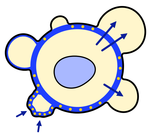
blebology.com is a public reference on cellular blebs, the rounded bulges that animal cells push out of their surface when the membrane comes away from the cortex beneath it.
It is written for scientists who are not bleb specialists, from cell biologists and physicists to engineers, clinician-scientists and students. We explain the basic concepts and go into enough depth that bleb specialists will also find it a useful resource.
What this site is
Blebs have been described for almost three centuries, under many names and in many organisms, but knowledge of them is scattered across cell biology, biophysics, developmental biology, cancer research and the study of cell death. This site brings it together and explains it, from how blebs form and retract to what remains unknown. It expands and updates the review “Blebology: principles of bleb-based migration”, by Juan Manuel García-Arcos, Ankita Jha, Clare M. Waterman and Matthieu Piel, published in Trends in Cell Biology in 2024 (García-Arcos et al., 2024a ◆), and is also an experiment in new ways of compiling and sharing scientific reviews. The pages rest on a systematic reading of the literature, described in the Meta-review, and every statement points to its source. We update the site as new work appears, discuss a recent paper each month in a journal club, and welcome corrections (About).
What is a bleb?
The surface of an animal cell has two layers. The plasma membrane lies on top of the cortex, a thin network of actin filaments pulled taut by myosin motors and attached to the membrane by linker proteins. Because the cortex is under tension, the cytoplasm it encloses is under pressure. A bleb forms where the membrane loses the support of the cortex, either because the links between them fail or because the cortex itself tears. The pressure then pushes the freed membrane outward, and cytoplasm flows in to fill a rounded protrusion within seconds (Charras et al., 2005; Tinevez et al., 2009). A new cortex assembles under the bleb membrane and stops its growth, and myosin then usually pulls the bleb back (Cunningham, 1995; Charras et al., 2006).
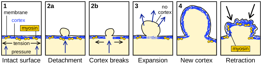
This simple cycle hides much variety. Some blebs last seconds and others hours; some stay in place, some travel around the cell, and some leave it altogether. Cells bleb when they migrate, divide and die, and blebs can also act as small compartments that organize signaling.
A guide to the site
How to read the pages
Each page reads as connected text, with details in the Milestones and in the reference list at the bottom, which opens with a click. Author–year citations link to the paper, and specialist terms link to the glossary at first use. Central claims carry a discreet italic label for the state of the evidence, from established to open question, explained on the About page. Our own papers are marked ◆.
References (5)
- Charras GT, Hu CK, Coughlin M et al. (2006) Reassembly of contractile actin cortex in cell blebs. J Cell Biol 175:477–490. doi:10.1083/jcb.200602085
- Charras GT, Yarrow JC, Horton MA et al. (2005) Non-equilibration of hydrostatic pressure in blebbing cells. Nature 435:365–369. doi:10.1038/nature03550
- Cunningham CC (1995) Actin polymerization and intracellular solvent flow in cell surface blebbing. J Cell Biol 129:1589–1599. doi:10.1083/jcb.129.6.1589
- ◆ García-Arcos JM, Jha A, Waterman CM et al. (2024a) Blebology: principles of bleb-based migration. Trends Cell Biol 34:838–853. doi:10.1016/j.tcb.2024.02.009
- Tinevez JY, Schulze U, Salbreux G et al. (2009) Role of cortical tension in bleb growth. Proc Natl Acad Sci USA 106:18581–18586. doi:10.1073/pnas.0903353106