M091982–1984 · Blebs on cultured animal cells, 1958–1984 · proposed
Blebs anchor lymphocytes moving through 3D matrix
In the early 1980s a crawling cell was thought to need firm contacts with its surroundings. Lymphocytes in collagen gels questioned this. Haston et al. (1982) proposed that their protrusions anchor the cell by their shape, wedged in gaps of the matrix, and Haston and Shields (1984) called the protrusions blebs and proposed as the drive a wave of contraction running backward along the cell through a stationary constriction ring. The mechanism remains proposed: it rests on attachment assays and kinematics, without perturbation of myosin, pressure or adhesion molecules.
Before
Stationary constrictions had been seen long before they were explained: De Bruyn (1946) filmed rabbit leukocytes passing rings at the same places along one path and blamed unseen obstacles. Senda et al. (1975) described in neutrophils on glass a contraction wave, a constriction formed at the front that stayed fixed relative to the substratum as the cell moved through it, and attributed it to the granuloplasm, the granular cytoplasm under the surface. Traction was thought to need adhesions (as stated by Haston et al., 1982), so Haston and colleagues asked whether this holds inside a three-dimensional (3D) matrix.
What was done
Haston et al. (1982) offered mouse lymph-node lymphocytes two-dimensional (2D) and 3D substrates of identical chemistry. The cells barely attached to glass coated with serum or fibronectin, attached better to hydrated than to dried collagen, and attached to filters with 3 and 8 µm pores but hardly to those with 0.22 and 0.45 µm pores (table): texture mattered more than composition. In collagen gels, protrusions expanded behind a constriction and stayed fixed while the cell body moved past them (three cells). The authors proposed adhesion-independent migration, with protrusions in gaps of the matrix as anchors; they called them pseudopodia and did not invoke pressure.
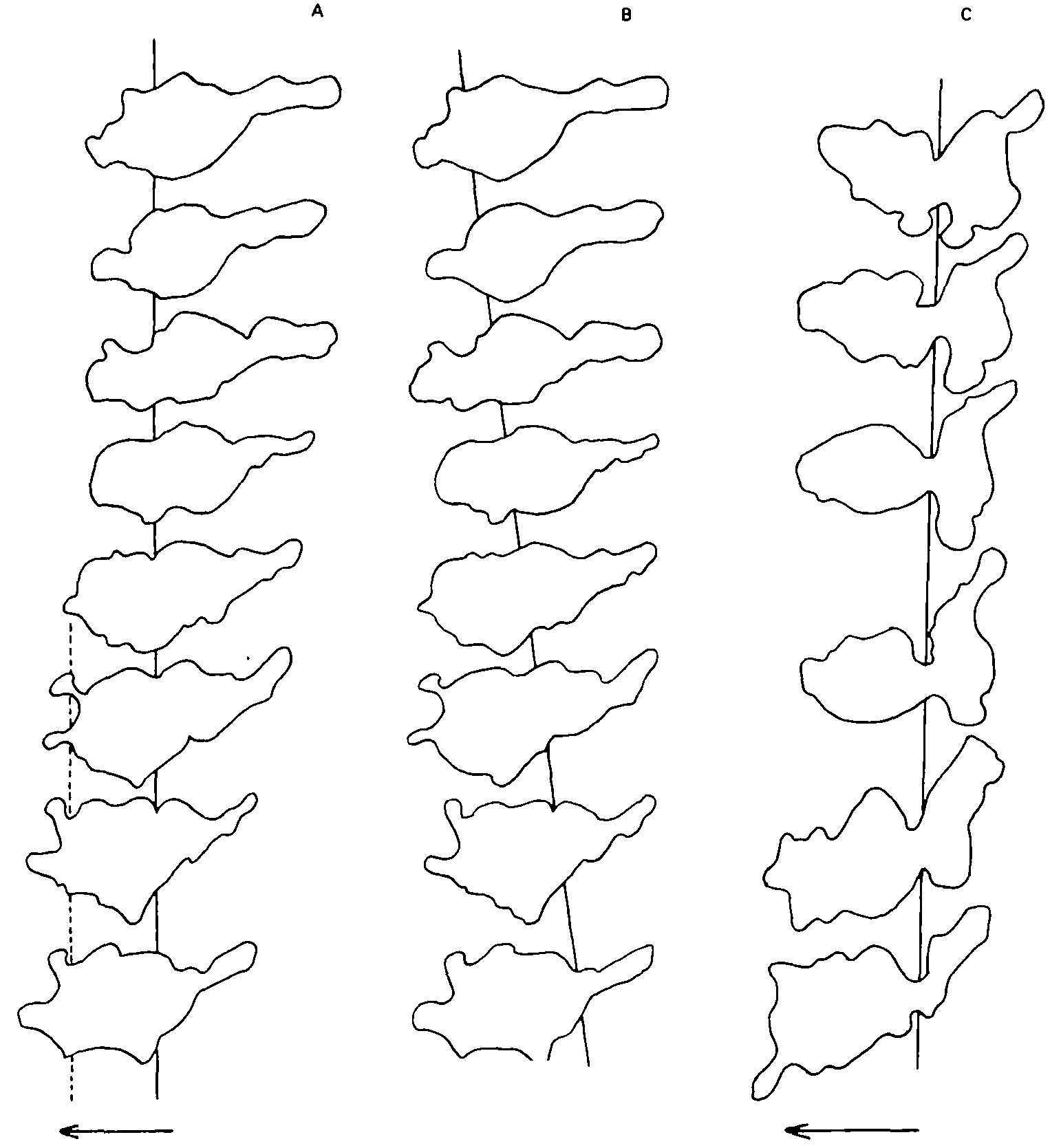
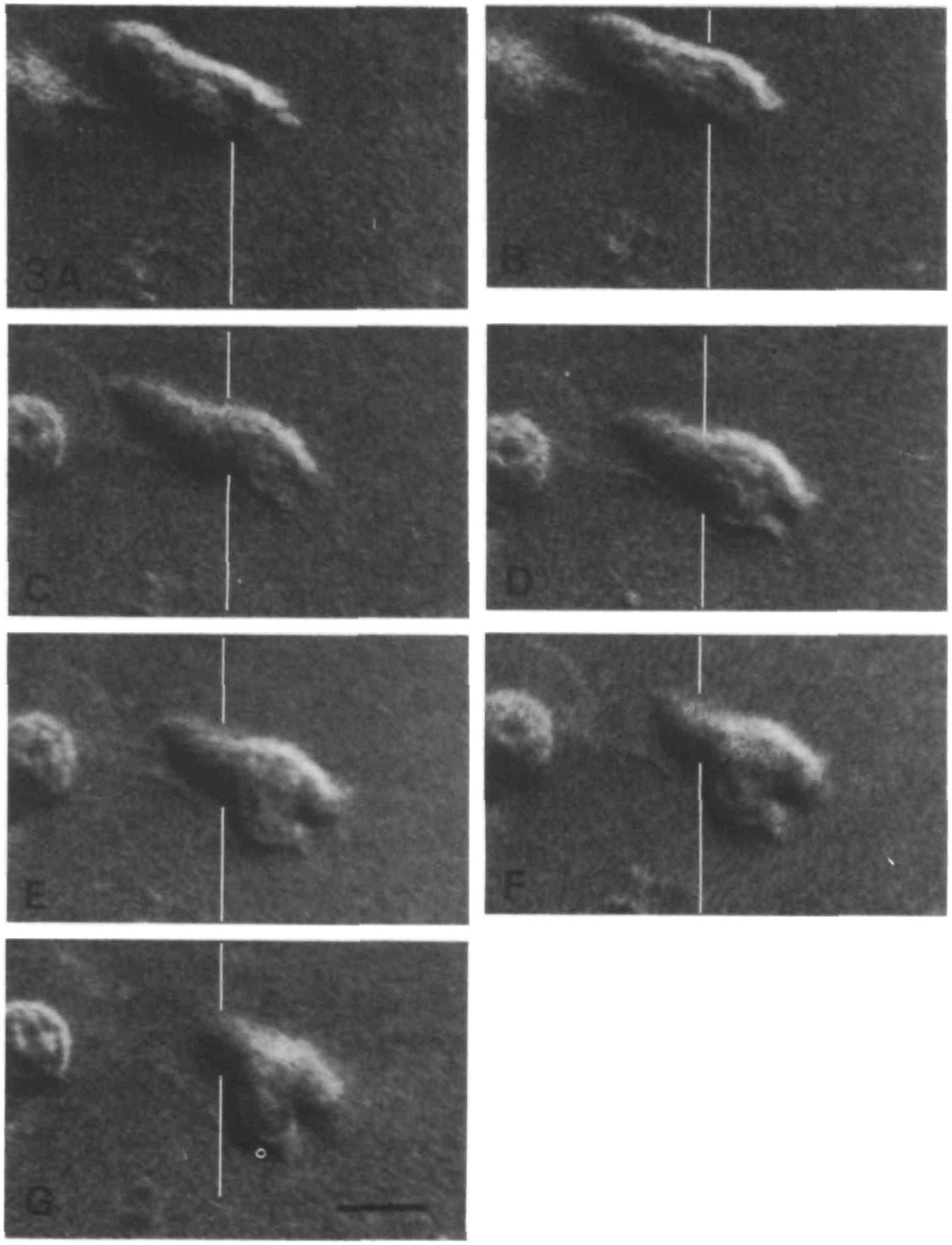
Haston and Shields (1984) then filmed mixed T and B cells every 2 or 4 s. In suspension, up to 80% of cells took a locomotor shape without moving, while a ring of contraction ran from front to back along each. In collagen the ring stayed fixed relative to the matrix, so in the cell’s frame it moved backward, and blebs expanded into the gel within 1–4 s and stayed in place as the cell body passed (figures). The same wave that produces no movement in suspension, the authors argued, moves the cell forward once its blebs are held by the gel. F-actin staining and electron microscopy showed nothing specialized at the ring.
They likened the movement to the peristalsis of worms with a hydrostatic skeleton: pressure would expand the anchored blebs, and pseudopod extension would be “an illusion created by the wave of contraction” (p. 240). In our reading, the new elements beyond Senda et al. (1975) were the wave in suspended lymphocytes, the 3D gel and the bleb anchors.
| Quantity | Value (units, error, n) | System | Source |
|---|---|---|---|
| Attachment to collagen coats, 37 °C | wet ≈52%, dried ≈15%, dried then rehydrated ≈35% of clean-glass binding (read from plot; no error bars) | mouse lymph-node lymphocytes, 3 mg/ml collagen, ⁵¹Cr label | Haston et al., 1982, Fig. 2 |
| Cells attached to serum-coated filters | 27 ± 8, 37 ± 11, 378 ± 161, 451 ± 112 at 0.22, 0.45, 3 and 8 µm pores (mean ± s.d., 4 fields × 2 filters) | same cells | Haston et al., 1982, Table II |
| Contraction-wave transit, suspension | mean 31 s (range 16–80 s; n = 54 cells) | mouse lymph-node lymphocytes, 37 °C, time-lapse | Haston and Shields, 1984, Fig. 1 |
| Passage through stationary constriction | mean 30 s (range 18–64 s; n = 59 cells) | 2.0 mg/ml collagen gel, time-lapse | Haston and Shields, 1984, Figs 2, 3 |
| Invasion front at 2 h vs adhesion to collagen-coated glass | 160–202 µm at 4–66% of clean-glass adhesion; 57 ± 8 µm at 110% (mean ± s.d., 15 readings, 3 gels) | rabbit peritoneal neutrophils | Brown, 1982, Table 1 |
What it changed
The two papers gave blebs a mechanical job, as anchors for movement through a 3D matrix without adhesions. The same year, rabbit neutrophils unable to move on collagen-coated glass were found to invade collagen gels, and invasion fell sharply only when Mn²⁺ raised their adhesion to 110% of that on clean glass (Brown, 1982; table). Brown proposed dilated pseudopodia as anchors, called the film evidence “not conclusive”, and did not mention blebs.
Later work tested the adhesion-free idea, not always with blebs. Leukocytes lacking all integrins moved through collagen at wild-type speed, by actin-driven protrusion and myosin II squeezing as that study proposed, though blebs were not imaged (Lämmermann et al., 2008). Anchoring by shape returned in a 2D model in which blebs interlocking with matrix gaps gave traction at zero adhesion (Tozluoğlu et al., 2013), and friction against a rearward cortical flow was shown to move confined, blebbing Walker carcinosarcoma cells (Bergert et al., 2015; M20).
Later work and open questions
Two caveats limit the reading of this work as bleb migration. Charras (2008) lists leukocytes among cells whose bleb-like fronts lacked demonstrated membrane detachment, and pressure was argued only by analogy with worms. Several leukocyte types also migrate without myosin II motor activity, which “largely excludes bleb formation” as their general mode (Renkawitz and Sixt, 2010, p. 747).
The constriction ring has had three readings: unseen obstacles (De Bruyn, 1946), which rings in suspended lymphocytes argue against; a contraction traveling back along the cell (Senda et al., 1975; Haston and Shields, 1984), for which Haston and Shields also cited Norberg (1971), although the ATP-driven ring he saw in one glycerol-killed lymphocyte formed in place and persisted; and, in blebbing Walker carcinosarcoma cells, the edge of a cortex left behind when the membrane detached (Keller and Eggli, 1998; M10). The cytoskeletal evidence does not decide between the last two: F-actin bands often sat at neutrophil rings in gels but not in suspension, uncounted (Haston, 1987), and a myosin ring traveled along suspended T-lymphoblasts (Bornens et al., 1989). In our reading, the two need not exclude each other, because they come from different cells.
Three questions remain open: whether lymphocyte blebs grip a fibrous matrix by shape, friction or residual adhesion, whether the ring is a traveling actomyosin wave or the edge of a detached cortex, and whether T cells in collagen use blebs at all.
References (14)
- Bergert M, Erzberger A, Desai RA et al. (2015) Force transmission during adhesion-independent migration. Nat Cell Biol 17:524–529. doi:10.1038/ncb3134
- Bornens M, Paintrand M, Celati C (1989) The cortical microfilament system of lymphoblasts displays a periodic oscillatory activity in the absence of microtubules: implications for cell polarity. J Cell Biol 109:1071–1083. doi:10.1083/jcb.109.3.1071
- Brown AF (1982) Neutrophil granulocytes: adhesion and locomotion on collagen substrata and in collagen matrices. J Cell Sci 58:455–467. doi:10.1242/jcs.58.1.455
- Charras GT (2008) A short history of blebbing. J Microsc 231:466–478. doi:10.1111/j.1365-2818.2008.02059.x
- De Bruyn PPH (1946) The amoeboid movement of the mammalian leukocyte in tissue culture. Anat Rec 95:177–191. doi:10.1002/ar.1090950209
- Haston WS (1987) F-actin distribution in polymorphonuclear leucocytes. J Cell Sci 88:495–501. doi:10.1242/jcs.88.4.495
- Haston WS, Shields JM (1984) Contraction waves in lymphocyte locomotion. J Cell Sci 68:227–241. doi:10.1242/jcs.68.1.227
- Haston WS, Shields JM, Wilkinson PC (1982) Lymphocyte locomotion and attachment on two-dimensional surfaces and in three-dimensional matrices. J Cell Biol 92:747–752. doi:10.1083/jcb.92.3.747
- Keller H, Eggli P (1998) Protrusive activity, cytoplasmic compartmentalization, and restriction rings in locomoting blebbing Walker carcinosarcoma cells are related to detachment of cortical actin from the plasma membrane. Cell Motil Cytoskeleton 41:181–193. doi:10.1002/(SICI)1097-0169(1998)41:2<181::AID-CM8>3.0.CO;2-H
- Lämmermann T, Bader BL, Monkley SJ et al. (2008) Rapid leukocyte migration by integrin-independent flowing and squeezing. Nature 453:51–55. doi:10.1038/nature06887
- Norberg B (1971) The formation of an ATP-induced constriction ring in a glycerinated lymphocyte during migration. Scand J Haematol 8:75–80. DOI not recorded.
- Renkawitz J, Sixt M (2010) Mechanisms of force generation and force transmission during interstitial leukocyte migration. EMBO Rep 11:744–750. doi:10.1038/embor.2010.147
- Senda N, Tamura H, Shibata N et al. (1975) The mechanism of the movement of leucocytes. Exp Cell Res 91:393–407. doi:10.1016/0014-4827(75)90120-2
- Tozluoğlu M, Tournier AL, Jenkins RP et al. (2013) Matrix geometry determines optimal cancer cell migration strategy and modulates response to interventions. Nat Cell Biol 15:751–762. doi:10.1038/ncb2775