M071958–1968 · Blebs on cultured animal cells, 1958–1984 · strongly supported
Blebs measured and modeled as pressure blow-outs
In the 1950s and 1960s blebs became objects of experiment, and in 1968 they received the earliest mathematical model of blebbing we found, in which internal pressure pushes the membrane out under Laplace’s law (Prothero and Spencer, 1968). That blebs are reversible, pressure-driven protrusions that expand fast and retract more slowly is strongly supported. The 1968 mechanism, with the membrane alone resisting the pressure, was proposed and later revised: the resisting tension is now placed in the actin cortex.
Before
Hughes (1952) had stated the idea in words: filming dividing cells, he saw blebs grow for a few seconds to 3–5 µm and shrink, usually more slowly, and suggested that cytoplasm escapes through a local weakness until the elastic tension of the bleb wall balances the cell’s hydrostatic pressure. He credited the first sighting to Peremeschko (1879), who saw dividing skin cells of newt larvae put out and withdraw ‘amoeboid processes’. Holtfreter (1946), in contrast, had concluded that the clear bulges circling isolated amphibian embryonic cells were not produced by local variations of internal pressure.
What was done
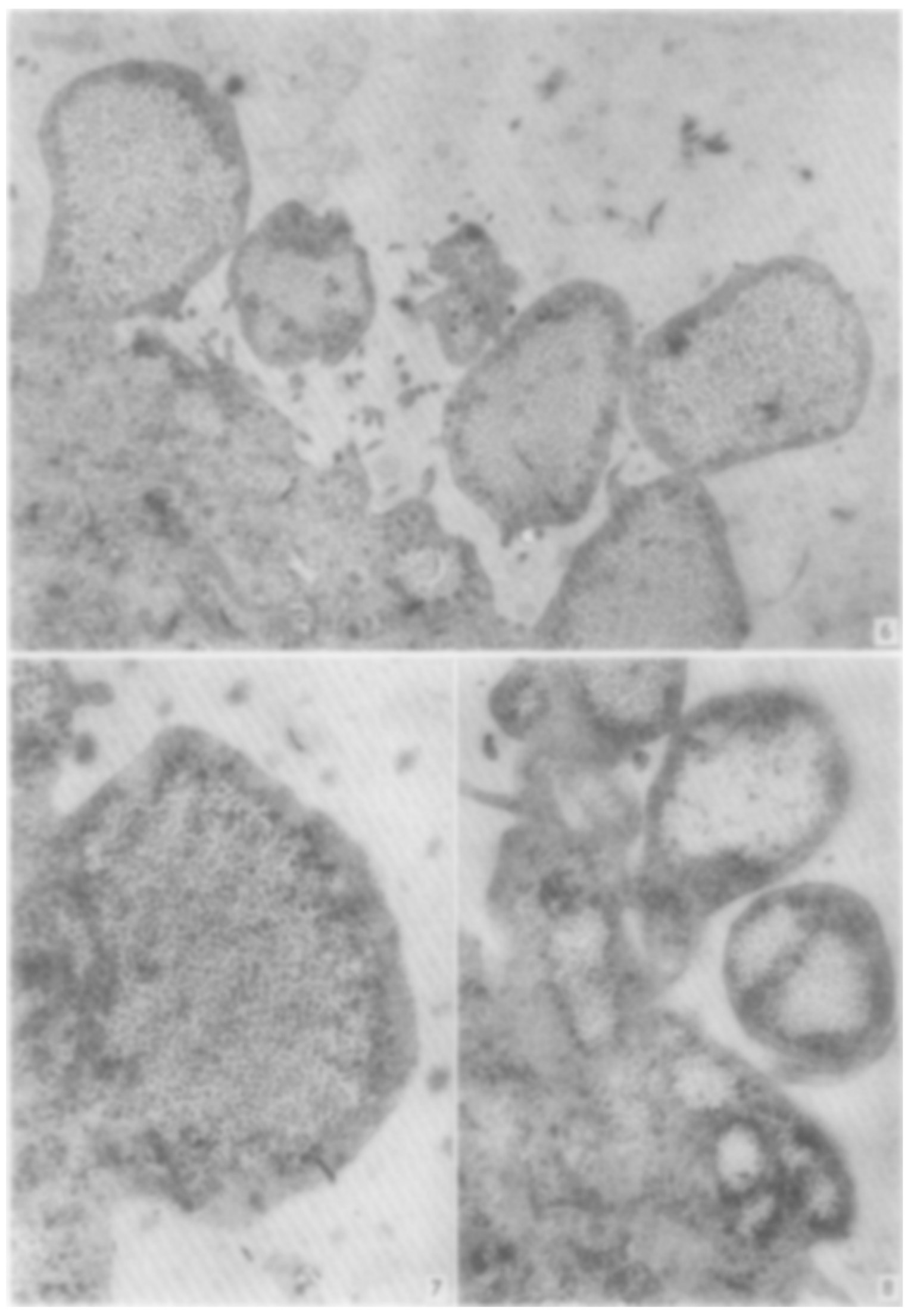
Dornfeld and Owczarzak (1958) made blebbing controllable. With the chelator EDTA, chick heart fibroblasts in 150 cultures contracted within 5 min and then blebbed continuously, recovering within 1–2 h of washout (table), whereas EDTA loaded with calcium, magnesium or iron had no effect. The authors proposed that unevenly rebuilt patches of the surface were “blown out” by hydrostatic pressure, an idea they called “admittedly speculative” (Dornfeld and Owczarzak, 1958, p. 248). In thin sections of one HEp cell, blebs held cytoplasmic matrix with endoplasmic reticulum or polysomes but no other organelles or fibers, with a dense rim under the membrane (Price, 1967; figure).
Prothero and Spencer (1968) filmed four mitotic epithelial cells from fetal mouse oviduct (table) and modeled them assuming a constant cell volume, an outward pressure from the contracting cleavage furrow and, centrally, a membrane whose tension stays constant as it is stretched over some range. They wrote “an equation of the form of LaPlace’s law” (Prothero and Spencer, 1968, p. 47):
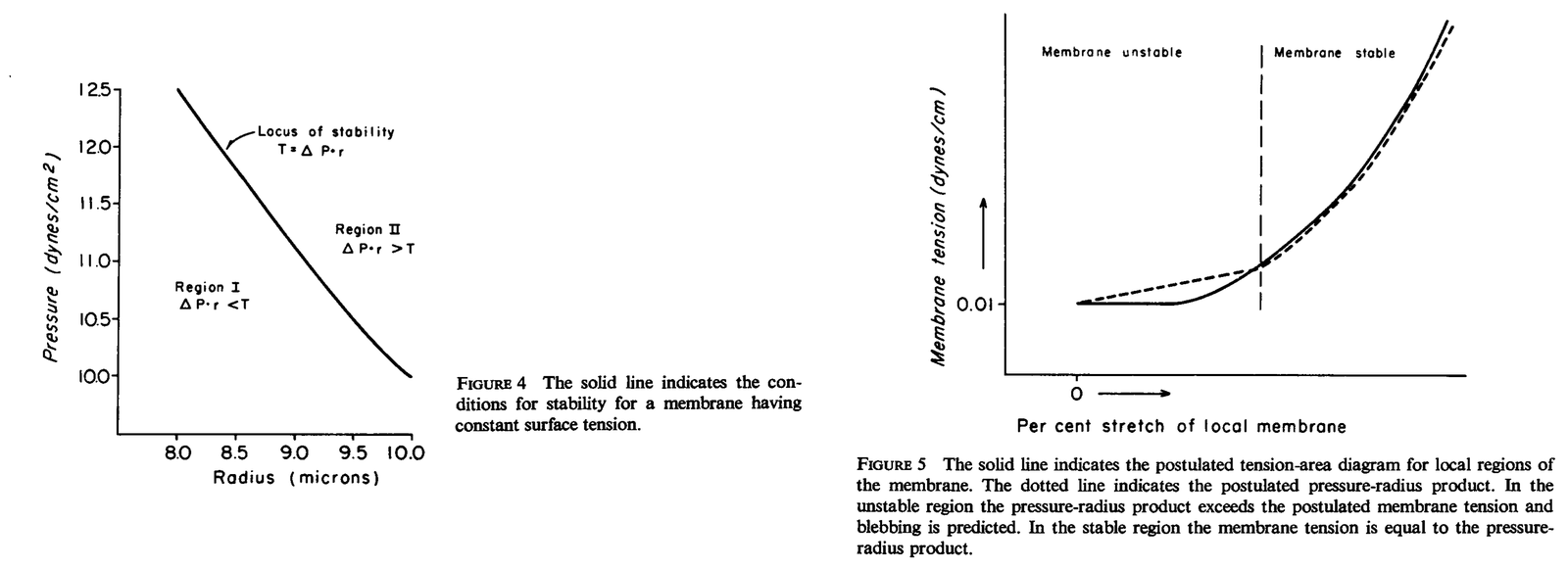
with \(T\) the membrane tension (0.01 dyn/cm = 10 pN/µm), \(\Delta P\) the excess intracellular pressure (10 dyn/cm² = 1 Pa) and \(r\) the radius of curvature (8–10 µm). A curved skin under tension holds a pressure difference across it, larger for a smaller radius. No geometry was named: this form holds for a cylinder, whereas a sphere gives \(\Delta P = 2T/r\) (Tinevez et al., 2009; our note). If the tension cannot rise, the surface is stable only while pressure times radius does not exceed it; beyond that, the membrane bulges until stretching raises the tension (figure). The values were assumed, not measured. The model also explained out-of-phase blebbing on opposite sides of a cell by conservation of volume, and the blebbing of dying cells by swelling.
| Quantity | Value | System | Source |
|---|---|---|---|
| Bleb diameter | 3.1 ± 1.0, 3.3 ± 0.5, 3.1 ± 0.7, 4.2 ± 0.8 µm (mean ± s.d.; n = 15, 15, 51, 18 blebs; 4 cells) | mitotic mouse oviduct epithelium, ≈30 °C, film | Prothero and Spencer, 1968, Table I |
| Rise time | 6.3 ± 1.9 and 5.5 ± 1.3 s (mean ± s.d.; 15 blebs in each of 2 cells) | same, continuous filming | Prothero and Spencer, 1968, Table I |
| Total duration | 30.7 ± 7.3 and 30.5 ± 7.1 s (continuous); 14.0 ± 2.8 and 18.1 ± 4.5 s (time-lapse) (mean ± s.d.) | same | Prothero and Spencer, 1968, Table I |
| Formation; withdrawal | ≈10 s; 20–30 s (n not stated) | chick heart fibroblasts, 0.003 M EDTA, time-lapse | Dornfeld and Owczarzak, 1958, text |
| Extension; retraction | ≤8 s; ≈1 min (average bleb; n not stated) | HEp cells, time-lapse | Price, 1967, text |
What it changed
All three studies found the asymmetry Hughes had seen, fast expansion and slower retraction, which later measurements confirmed: 4–7 s of expansion against about 30 s of retraction in melanoma cells (Dai and Sheetz, 1999), and a similar pattern for laser-induced blebs in L929 cells (Tinevez et al., 2009). In cytokinesis, Sedzinski et al. (2011) blebbed the contracting pole of oscillating HeLa cells by laser ablation, which arrested and reversed the flow between the poles (15 of 16 cells), and proposed blebs as pressure sinks without measuring pressure. Later work kept pressure and Laplace’s law but moved the resisting tension into the actomyosin cortex (Miranda et al., 1974a; Tinevez et al., 2009; M08).
Later work and open questions
The numbers show why the tension had to move. The assumed membrane tension is close to the 11–12 pN/µm later derived from tethers pulled from melanoma blebs, but the assumed pressure is far below later estimates, 20.7 ± 5.9 Pa in the same cells (mean ± s.d.; Dai and Sheetz, 1999) and 45–300 Pa in metaphase HeLa cells (Charras et al., 2008). Balancing tens to hundreds of pascals at a radius of about 10 µm needs a much larger tension, such as the cortical tension of about 400 pN/µm measured by aspiration in L929 cells (Tinevez et al., 2009; our arithmetic).
Constant volume holds (Tinevez et al., 2009), but dying cells need not swell to bleb: ATP-depleted hepatocytes blebbed whether their volume rose or fell (Gores et al., 1989). Whether pressure is shared across the cell, as the volume argument assumed, is still debated: it did not equilibrate over about 10 µm and 10 s in M2 melanoma cells (Charras et al., 2005), whereas in L929 cells a second bleb was equally smaller near or far from the first (Tinevez et al., 2009), with different cells, perturbations and timescales (M12).
The data are thin: four filmed cells, one sectioned cell and no counts for the EDTA experiments. Do polar blebs act by releasing pressure? Is Price’s dense rim, similar in thickness to the ≈50 nm cortex of retracting blebs (Charras et al., 2008), an early cortex, although expanding blebs show no actin enrichment (Charras et al., 2005; our comparison)? And what sets the ratio of retraction to expansion time, about 2 to 8 across these systems (open question)?
References (13)
- Charras GT, Yarrow JC, Horton MA et al. (2005) Non-equilibration of hydrostatic pressure in blebbing cells. Nature 435:365–369. doi:10.1038/nature03550
- Charras GT, Coughlin M, Mitchison TJ et al. (2008) Life and times of a cellular bleb. Biophys J 94:1836–1853. doi:10.1529/biophysj.107.113605
- Dai J, Sheetz MP (1999) Membrane tether formation from blebbing cells. Biophys J 77:3363–3370. doi:10.1016/S0006-3495(99)77168-7
- Dornfeld EJ, Owczarzak A (1958) Surface responses in cultured fibroblasts elicited by ethylenediaminetetraacetic acid. J Biophys Biochem Cytol 4:243–250. doi:10.1083/jcb.4.3.243
- Gores GJ, Flarsheim CE, Dawson TL et al. (1989) Swelling, reductive stress, and cell death during chemical hypoxia in hepatocytes. Am J Physiol 257:C347–C354. doi:10.1152/ajpcell.1989.257.2.C347
- Holtfreter J (1946) Structure, motility and locomotion in isolated embryonic amphibian cells. J Morphol 79:27–62. doi:10.1002/jmor.1050790103
- Hughes A (1952) The Mitotic Cycle: the Cytoplasm and Nucleus during Interphase and Mitosis. Butterworths, London. No DOI.
- Miranda AF, Godman GC, Deitch AD et al. (1974a) Action of cytochalasin D on cells of established lines. I. Early events. J Cell Biol 61:481–500. doi:10.1083/jcb.61.2.481
- Peremeschko (1879) Ueber die Theilung der thierischen Zellen. Arch Mikrosk Anat 16:437–457. doi:10.1007/bf02956388
- Price ZH (1967) The micromorphology of zeiotic blebs in cultured human epithelial (HEp) cells. Exp Cell Res 48:82–92. doi:10.1016/0014-4827(67)90278-9
- Prothero JW, Spencer D (1968) A model of blebbing in mitotic tissue culture cells. Biophys J 8:41–51. doi:10.1016/S0006-3495(68)86473-2
- Sedzinski J, Biro M, Oswald A et al. (2011) Polar actomyosin contractility destabilizes the position of the cytokinetic furrow. Nature 476:462–466. doi:10.1038/nature10286
- Tinevez JY, Schulze U, Salbreux G et al. (2009) Role of cortical tension in bleb growth. Proc Natl Acad Sci USA 106:18581–18586. doi:10.1073/pnas.0903353106