M162023 · Blebs as signaling hubs · strongly supported
Blebs as curvature-based signaling hubs for survival
Blebs can do more than move cells. In melanoma cells kept without adhesion, septins, cytoskeletal proteins that recognize micron-scale membrane curvature (Bridges et al., 2016), gathered at the bases of dynamic blebs, where the membrane, seen from inside the cell, curves positively. There they formed a signaling hub that held the RAS protein NRAS and kept ERK and PI3K signaling active, so that blebbing protected the cells from anoikis, the death of cells deprived of adhesion (Weems et al., 2023). This chain is strongly supported in cell lines, from one laboratory; the test in animals is a preprint.
Before
Two older observations prepared this result. The first is that cells bleb when they lose adhesion, but only for a while. Trypsin-detached CHO cells went from 62% blebbing at 6 min to 10% at 40 min, as blebs gave way to microvilli (Kinn and Allen, 1981), and detached endothelial cells stopped within an hour unless treated with the dynamin inhibitor dynasore (Norman et al., 2010). In re-plated HeLa and CHO cells, blebs held active FAK and phosphorylated MAPK and were proposed, from images alone, as transient signaling platforms (Moes et al., 2011).
The second is that blebbing can help injured cells survive. After damage by the pore-forming toxin streptolysin O, 82 ± 6% of blebbing HEK 293 cells recovered against 38 ± 6% of non-blebbing ones (mean ± s.e.m.; n = 85 and 140 cells), and in example cells calcium entry and loss of cytoplasm were confined to single blebs (Babiychuk et al., 2011).
What was done
Weems et al. (2023) compared detached cells with adherent controls, using three melanoma lines, MV3 with an NRAS mutation and A375 and M498 with a BRAF mutation, and mouse embryonic fibroblasts. Do detached cells need their blebs? Wheat germ agglutinin (WGA), which reduces blebbing (Charras et al., 2008), killed detached but few adherent MV3 cells, and cells held in non-adhesive hydrogels behaved like detached ones.
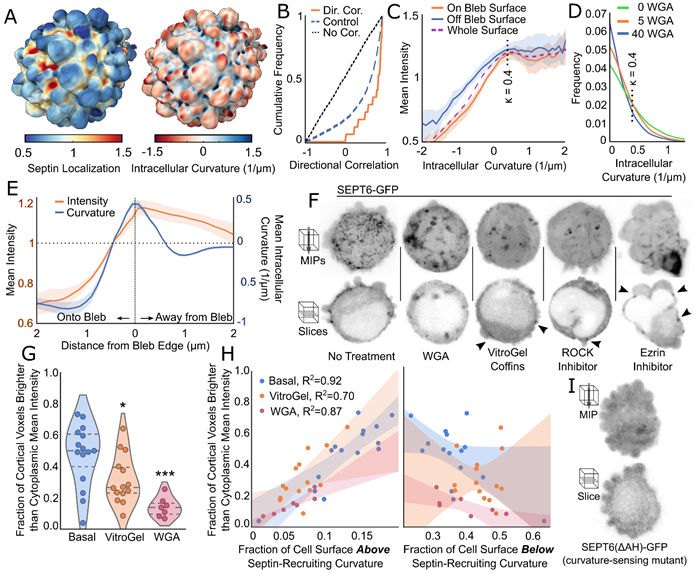
How, then, could blebs send a survival signal? Seen from inside the cell, membrane curvature is negative on a bleb and positive at its base. Tagged septin 6 rose with positive curvature up to a plateau and peaked at bleb edges, every manipulation that reduced blebbing also lowered cortical septin, and a septin 6 lacking its curvature-sensing amphipathic helix (Cannon et al., 2019) stayed off the surface. In this picture, blebs create the curvature that septins read.
The rest of the chain rests on perturbations that separate septins from blebs. The septin inhibitor forchlorfenuron removed septin from the cortex without changing blebbing, yet it killed detached, bleb-dependent cells. Among 321 septin partners found by proximity labeling was NRAS, which lost its cortical enrichment when septins were inhibited, and a dominant-negative NRAS killed detached cells. Inhibiting septins or blebs lowered ERK activity in detached but not adherent cells, and PI3K activity in rounded cells in collagen. Conversely, dominant-negative dynamin, chosen because of the dynasore result, kept detached fibroblasts blebbing and lowered caspase activation, an effect that WGA or forchlorfenuron undid.
| Quantity | Value | System | Source |
|---|---|---|---|
| Cortical septin against surface fraction with curvature > 0.4 µm⁻¹ | R² = 0.92 / 0.70 / 0.87 (basal / hydrogel / WGA) | MV3, septin 6–GFP | Weems et al., 2023, Fig. 2H |
| Extra death with WGA 20 µg/ml, detached vs adherent | ≈0.5 vs ≈0.17 (read from plot; 95% CI; 3 paired experiments; 426–688 cells per dose) | MV3, 24 h | Weems et al., 2023, Fig. 1B |
| ERK activity change with forchlorfenuron, detached vs adherent | ≈ −25 to −58% vs ≈ −6 to +7% (4 experiments; 234–405 cells per group; read from plot) | MV3, 3 h | Weems et al., 2023, Fig. 4E |
| Extra caspase-active fraction, detached minus adherent | ≈0.07–0.17 with dominant-negative dynamin (prolonged blebbing) vs ≈0.16–0.48 control (3 replicates) | mouse embryonic fibroblasts, 4 h | Weems et al., 2023, Fig. 5C |
What it changed
Commentaries presented blebs as signaling platforms (Reichman-Fried and Raz, 2023; Strzyz, 2023), and an independent group reached a related result by another route. In detached MDA-MB-231 cells blebbing under latrunculin B, PI(3,4,5)P3 was higher and PI(4,5)P2 lower on bleb than on non-bleb membrane, the adaptor SH3KBP1 bridged septin 6 and a PI3K subunit at the bleb neck, and blocking blebs, septins, PI3K or SH3KBP1 raised cell death (Zhen et al., 2025). Because the blebs were drug-induced and their number was never measured, this support is limited.
The Weems group then looked in tissue (Weems et al., 2026 preprint). Rounded, blebbing cells in patient samples, mostly melanoma, and at the invasive margins of tumors showed septin enriched at the membrane, judged by eye. In MV3 xenografts, oral forchlorfenuron raised cell death at the tumor margin and lowered recurrence after surgical removal from 66% to 23% (25 mice). More is on Blebs as signaling hubs.
Later work and open questions
Beyond the usual limits of work in cell lines, such as inhibitors that may act off target, overexpressed septin and NRAS, and readouts from two to four experiments, the curvature argument has two weak points. On a rugged three-dimensional surface, even a uniform membrane label looks enriched at bleb edges (Driscoll et al., 2019), which Weems et al. answered mainly with the helix mutant. And none of the three in vitro sources cited for their curvature threshold of 0.4 µm⁻¹ names a threshold: 0.4 µm⁻¹ was the shallowest bead tested (Cannon et al., 2019), a bead that bound little or no septin (Bridges et al., 2016), or a point on a gradual rise of septin density toward a maximum near +2 µm⁻¹ (Beber et al., 2019). Lipid data also differ between systems: PI(4,5)P2 was higher on spontaneous MV3 blebs (Driscoll et al., 2019) but depleted from latrunculin-induced blebs (Zhen et al., 2025).
The in vivo preprint infers blebs from fixed morphology and perturbs only septins, which also act away from blebs, so it tests the hub rather than the bleb. Whether blebs themselves support the survival of disseminating cells in tissue, and whether other blebbing cells build such hubs, is not known, so blebs as signaling hubs in general remain proposed.
References (14)
- Babiychuk EB, Monastyrskaya K, Potez S et al. (2011) Blebbing confers resistance against cell lysis. Cell Death Differ 18:80–89. doi:10.1038/cdd.2010.81
- Beber A, Taveneau C, Nania M et al. (2019) Membrane reshaping by micrometric curvature sensitive septin filaments. Nat Commun 10:420. doi:10.1038/s41467-019-08344-5
- Bridges AA, Jentzsch MS, Oakes PW et al. (2016) Micron-scale plasma membrane curvature is recognized by the septin cytoskeleton. J Cell Biol 213:23–32. doi:10.1083/jcb.201512029
- Cannon KS, Woods BL, Crutchley JM et al. (2019) An amphipathic helix enables septins to sense micrometer-scale membrane curvature. J Cell Biol 218:1128–1137. doi:10.1083/jcb.201807211
- Charras GT, Coughlin M, Mitchison TJ et al. (2008) Life and times of a cellular bleb. Biophys J 94:1836–1853. doi:10.1529/biophysj.107.113605
- Driscoll MK, Welf ES, Jamieson AR et al. (2019) Robust and automated detection of subcellular morphological motifs in 3D microscopy images. Nat Methods 16:1037–1044. doi:10.1038/s41592-019-0539-z
- Kinn SR, Allen TD (1981) Conversion of blebs to microvilli: cell surface reorganisation after trypsin. Differentiation 20:168–173. doi:10.1111/j.1432-0436.1981.tb01172.x
- Moes MJ, Bijvelt JJ, Boonstra J (2011) Attachment of HeLa cells during early G1 phase. Histochem Cell Biol 136:399–411. doi:10.1007/s00418-011-0852-9
- Norman LL, Brugués J, Sengupta K et al. (2010) Cell blebbing and membrane area homeostasis in spreading and retracting cells. Biophys J 99:1726–1733. doi:10.1016/j.bpj.2010.07.031
- Reichman-Fried M, Raz E (2023) Bleb protrusions help cancer cells to cheat death. Nature 615:402–403. doi:10.1038/d41586-023-00477-4
- Strzyz P (2023) Anoikis resistance through blebs. Nat Rev Mol Cell Biol 24:310. doi:10.1038/s41580-023-00598-8
- Weems AD, Welf ES, Driscoll MK et al. (2023) Blebs promote cell survival by assembling oncogenic signalling hubs. Nature 615:517–525. doi:10.1038/s41586-023-05758-6
- Weems AD, Islam MT, Borges H et al. (2026) In vivo evidence for bleb-induced survival signaling in metastatic melanoma. bioRxiv. doi:10.64898/2026.01.05.697601 preprint
- Zhen Z, Zhang C, Li J et al. (2025) Blebs regulate phosphoinositide distribution and promote cell survival through the Septin-SH3KBP1-PI3K axis. Am J Physiol Cell Physiol 329:C145–C158. doi:10.1152/ajpcell.00096.2025