M142016–2024 · Blebs as physical objects · strongly supported
Inverse blebs: pressure from outside the cell
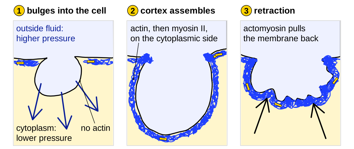
A bleb normally bulges outward because the cytoplasm is at higher pressure than the medium. Work since 2016 showed that the cycle can run in reverse: where the fluid outside a cell is at higher pressure, the membrane bulges into the cell without actin beneath it, and actomyosin then pulls it back. These inverse blebs take part in forming lumens in zebrafish vessel sprouts and in the 32-cell mouse embryo (Gebala et al., 2016; Schliffka et al., 2024), and they also form in bile canaliculi of the liver (Gupta et al., 2017). Pressure-dependent inverse blebs retracted by myosin II are strongly supported in all three systems; their role as hydraulic pumps is proposed, because neither pressure nor fluid flow was measured.
Before
In the standard account, cortical contraction pressurizes the cytoplasm, which pushes out membrane that has detached from the cortex or lacks it (M12). Nothing in this account fixes the sign of the pressure difference, yet we found no description of an inward bleb before 2016. Two problems of lumen formation made the question relevant. In growing blood vessels, lumens were thought to form without flow, by fusion of vacuoles inside endothelial cells (as summarized by Gebala et al., 2016). In the mouse embryo, pressurized fluid, which the authors traced to the medium, fractures the contacts between cells into hundreds of small pockets, or microlumens, that later merge into one cavity; blocking the sodium pump prevented the pockets (Dumortier et al., 2019). How fluid moves between pockets was unclear.
What was done
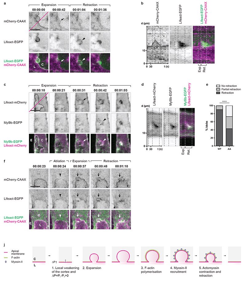
Gebala et al. (2016) imaged intersegmental vessels, sprouts that grow from the dorsal aorta of zebrafish embryos. The luminal (apical) membrane of the endothelial cells bulged spherically into the cells, a process the authors named ‘inverse membrane blebbing’, and no vacuoles were seen. Stopping blood flow reversibly with tricaine, or cutting the sprout from the aorta, stopped blebbing, and the lumen regressed (5–7 embryos per test), so the authors inferred that blood pressure pushes the membrane inward. The blebs expanded free of F-actin and myosin II, which then assembled in that order before retraction, as on outward blebs (M13; figure). A laser cut in the apical cortex produced an inverse bleb, and a myosin light chain that cannot be phosphorylated impaired retraction and lumen formation (table).
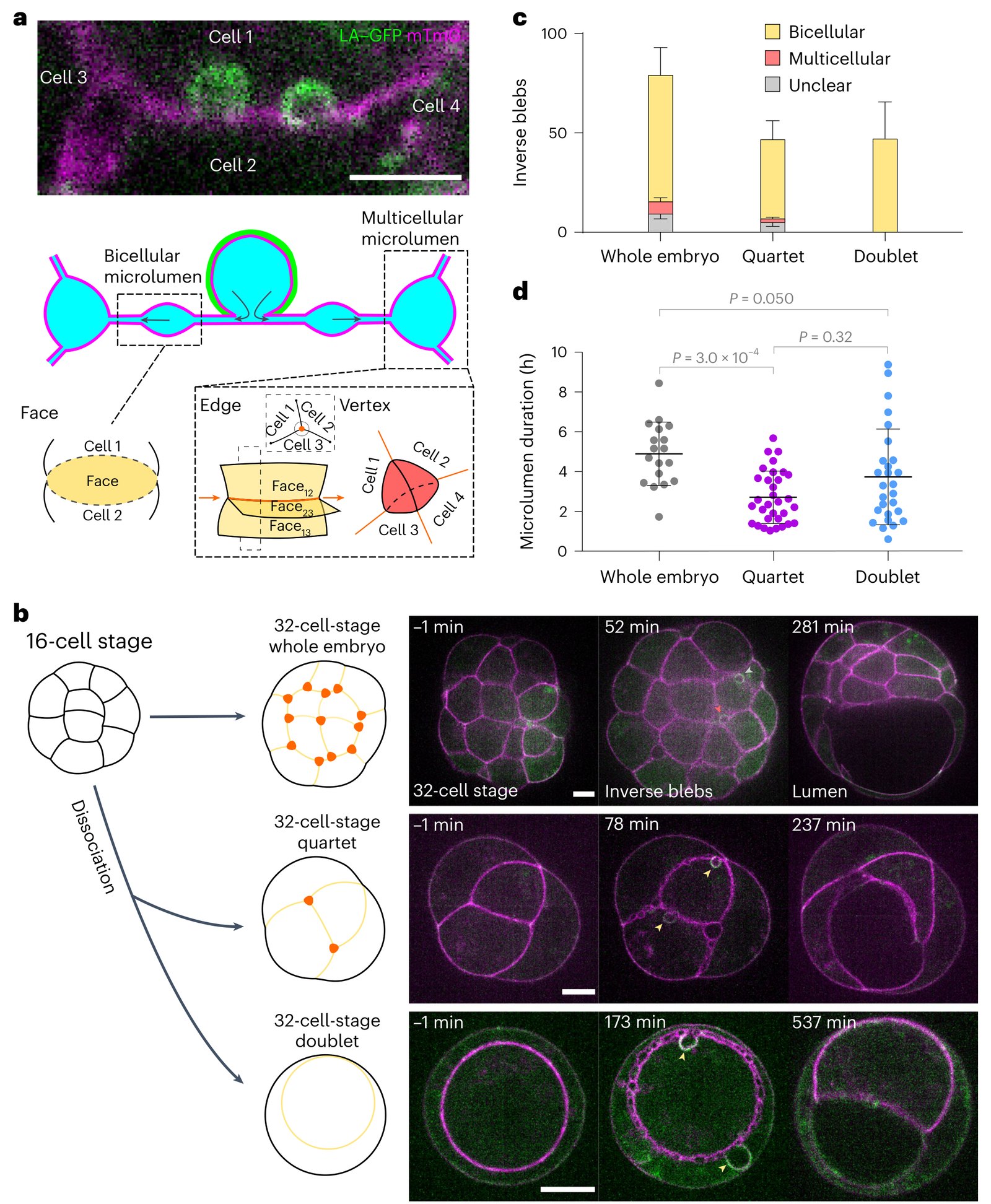
Schliffka et al. (2024) found the same cycle at contacts between cells of 32-cell mouse embryos. Hemispherical intrusions grew several micrometers into cells within about 10 s and filled with dextran from the fluid between cells, which identified them as pockets of that fluid rather than outward blebs of a neighbor. Actin appeared shortly before maximal size and myosin about 15 s later. Blebbing coexisted with microlumens and depended on adhesion and fluid transport: maternal loss of E-cadherin strongly reduced it, and EIPA, which blocks sodium transport, almost abolished it. Reducing myosin II slowed retraction without changing bleb size. The authors proposed that the blebs pump fluid between microlumens and tested the idea by changing the topology of the embryo (figure).
| Quantity | Value | System | Source |
|---|---|---|---|
| Lateral blebs fully retracted, wild type vs Myl9bAA (non-phosphorylatable myosin light chain) | ≈90% vs ≈43% (read from plot; 102 vs 161 blebs, 5 cells each; p = 2.1 × 10⁻¹³) | zebrafish intersegmental vessel, 34 h post-fertilization | Gebala et al., 2016, Fig. 4e |
| Vessels with normal lumen, wild type vs Myl9bAA | ≈82% vs ≈53% (read from plot; 55 vessels/24 embryos vs 31/9; p = 0.00024) | zebrafish, 2 days post-fertilization | Gebala et al., 2016, Fig. 5a |
| Bleb size; expansion; retraction speed | 3.4 ± 4 µm; 0.08 ± 0.04; 0.03 ± 0.01 µm/s (mean ± s.d., 31 blebs, 3 cells) | zebrafish intersegmental vessel | Gebala et al., 2016, Supplementary Fig. 1e |
| Embryos with inverse blebs, wild type vs maternal Cdh1+/− (E-cadherin) | 18/18 vs 5/15 (P = 3.3 × 10⁻⁵) | 32-cell mouse embryo | Schliffka et al., 2024, Extended Data Fig. 4c |
| Retraction time, wild type / maternal Myh9+/− / Myh10+/− | ≈36 / ≈46 / ≈34 s (read from plot; s.d. ≈11–14 s; 100 / 67 / 41 blebs; P = 1.3 × 10⁻⁵, 0.81; the text gives ≈50 s, measured differently) | 32-cell mouse embryo | Schliffka et al., 2024, Fig. 4b |
What it changed
Gebala et al. (2016) concluded that blebbing does not need a particular membrane polarity, and proposed a pressure-driven, myosin-limited bleb cycle as an alternative to vacuole fusion in shaping vessel lumens. In mouse liver, bile canaliculi, the fine channels into which liver cells secrete bile, formed inward blebs 1–2 µm across that lasted about 2 min; in cultured hepatocytes they began at local ruptures of the cortex and recruited actin and ezrin, then myosin IIA. The authors linked them to the backflow of bile in obstructive cholestasis rather than to lumens (Gupta et al., 2017). In our reading, these studies make the sign of the pressure difference a free parameter of bleb physics: wherever the outside is at higher pressure and the cortex is locally weak, the cycle can run inward (proposed).
Later work and open questions
The main limitation is that pressure was inferred, never measured: in vessels from stopping the heart and cutting the vessel, which also change other properties of the tissue; in the liver from geometry and drugs; in the embryo from adhesion loss and blocked sodium transport, without tracking fluid (Gebala et al., 2016; Gupta et al., 2017; Schliffka et al., 2024). The pump test also has weak points: reduced embryos of four and of two cells did not differ significantly (P = 0.32), slower retraction left the timing of microlumen coarsening unchanged, and bleb positions did not predict where the lumen formed. Replication is limited: the vessel work was extended by a group including a co-author of Gebala et al. (Kondrychyn et al., 2020), and the embryo work comes from one laboratory.
How an inverse bleb starts may differ between tissues. In vessels, cortex ablation suggests that local weakening is enough, as for outward blebs; in the embryo, the authors proposed that pressure opens a pocket bounded by adhesion between two cells (Schliffka et al., 2024). How much fluid one cycle moves, and how widespread inverse blebs are, remain open; giant vacuoles in Schlemm’s canal have been modeled as one candidate (Cairoli et al., 2023). In work on cell death, ‘inward blebbing’ means something unrelated, the uptake of plasma membrane into clear vesicles (Uchiyama, 1995).
References (8)
- Cairoli A, Spenlehauer A, Overby DR, Lee CF (2023) Model of inverse bleb growth explains giant vacuole dynamics during cell mechanoadaptation. PNAS Nexus 2:pgac304. doi:10.1093/pnasnexus/pgac304
- Dumortier JG, Le Verge-Serandour M, Tortorelli AF et al. (2019) Hydraulic fracturing and active coarsening position the lumen of the mouse blastocyst. Science 365:465–468. doi:10.1126/science.aaw7709 (bioRxiv preprint, doi:10.1101/537985, superseded)
- ◆ García-Arcos JM, Jha A, Waterman CM et al. (2024a) Blebology: principles of bleb-based migration. Trends Cell Biol 34:838–853. doi:10.1016/j.tcb.2024.02.009
- Gebala V, Collins R, Geudens I et al. (2016) Blood flow drives lumen formation by inverse membrane blebbing during angiogenesis in vivo. Nat Cell Biol 18:443–450. doi:10.1038/ncb3320
- Gupta K, Li Q, Fan JJ et al. (2017) Actomyosin contractility drives bile regurgitation as an early response during obstructive cholestasis. J Hepatol 66:1231–1240. doi:10.1016/j.jhep.2017.01.026 (bioRxiv 2016 preprint, doi:10.1101/077792, superseded)
- Kondrychyn I, Kelly DJ, Carretero NT et al. (2020) Marcksl1 modulates endothelial cell mechanoresponse to haemodynamic forces to control blood vessel shape and size. Nat Commun 11:5476. doi:10.1038/s41467-020-19308-5
- Schliffka MF, Dumortier JG, Pelzer D et al. (2024) Inverse blebs operate as hydraulic pumps during mouse blastocyst formation. Nat Cell Biol 26:1669–1677. doi:10.1038/s41556-024-01501-z (earlier version: bioRxiv 2023, doi:10.1101/2023.05.03.539105 preprint, superseded)
- Uchiyama Y (1995) Apoptosis: the history and trends of its studies. Arch Histol Cytol 58:127–137. doi:10.1679/aohc.58.127