M132006–2010 · Blebs as physical objects · strongly supported
Cortex reassembly and membrane–cortex attachment control blebbing
Once a bleb was understood as membrane freed from the cortex (M10), work in 2006–2010 showed how the cortex returns and what holds it to the membrane. A retracting bleb rebuilds its cortex in a fixed order, from the ERM proteins (ezrin, radixin and moesin), which link membrane to actin, to myosin II (Charras et al., 2006), and the energy of membrane–cortex attachment, set largely by ERM proteins, controls how often and how large cells bleb, in culture and in the zebrafish embryo (Diz-Muñoz et al., 2010). Both findings are strongly supported, with one apparent exception.
Before
In M2 melanoma cells, phalloidin stained fully extended but not expanding blebs, and blebs on cells treated with cytochalasin D never retracted; Cunningham (1995) proposed that expansion stops when local actin polymerization catches up. Tethers pulled from M2 cells showed that attachment to the cortex supplies most of the membrane’s apparent tension, and Dai and Sheetz (1999) proposed that a bleb forms where pressure exceeds this binding. Missing were the order of assembly and a test of attachment in an organism.
What was done
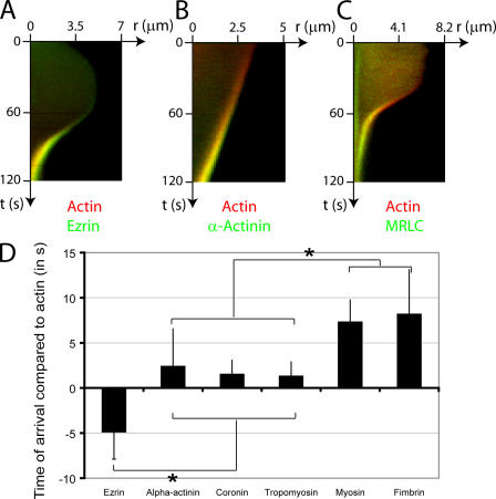
Charras et al. (2006) used the constant blebbing of M2 cells as a supply of membrane without cortex. They timed the arrival of cortical proteins at the bleb rim against actin. Expanding blebs carried no detectable actin. As expansion stopped, ezrin and moesin arrived first, about 5 s before actin (table); α-actinin, coronin and tropomyosin-4 followed actin within seconds, and fimbrin and the myosin regulatory light chain came last, completing a contractile cortex in about 30 s (figure).
Perturbations assigned roles. Cytochalasin D blocked recruitment of actin but not of ezrin, which therefore reaches the membrane independently. When an injected ezrin fragment uncoupled cellular ezrin from actin, the new actin rim tore away from the membrane as retraction began, whereas injected active ezrin almost stopped blebbing (5% of 21 cells against 54% of 79 controls). Under blebbistatin, myosin arrived but blebs did not retract. The authors concluded that ezrin “played no role in actin nucleation, but was essential for tethering” (p. 477), and inferred, without measuring it, that ezrin sets membrane–actin adhesion energy.
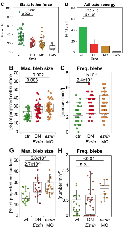
Diz-Muñoz et al. (2010) measured that energy and tested its role in an embryo. Membrane tethers pulled with an atomic force microscope from isolated progenitor cells of the zebrafish prechordal plate give an apparent tension equal to the membrane tension plus an attachment energy per unit area, W0. Reducing ERM function lowered W0 three- to fourfold without changing cortex tension, and isolated cells with weaker attachment formed larger and more frequent blebs (table, figure). In the embryo, ERM loss enlarged blebs, raised their share of leading-edge protrusions and made migration less persistent. The authors concluded that attachment and intracellular pressure together control bleb formation.
| Quantity | Value | System | Source |
|---|---|---|---|
| Ezrin arrival relative to actin | −4.87 ± 3 s (s.d., n = 23 blebs) | M2 melanoma blebs | Charras et al., 2006, Fig. 5D |
| Myosin regulatory light chain / fimbrin | +7.3 ± 2.4 s (n = 27) / +8.17 ± 4.9 s (n = 17) (s.d.) | M2 blebs | Charras et al., 2006, Fig. 5D |
| Attachment energy density W0 | ≈46 (± ≈15, abs. deviation of median) vs ≈16 (dominant-negative), ≈12 (morpholino) × 10⁻¹⁸ J/µm² (read from plot); tether force medians 32, 19.1, 17 pN from 114/56, 48/24, 76/45 tethers/cells | isolated zebrafish progenitors, atomic force microscope tethers | Diz-Muñoz et al., 2010, Fig. 1D; Table S1B |
| Maximum bleb size in vivo | ≈16% vs ≈24% (dominant-negative), ≈27% (morpholino) of projected cell area (medians; n = 14, 16, 15 blebs) | prechordal plate, 8–10 h post-fertilization | Diz-Muñoz et al., 2010, Fig. 2G |
| Later: ezrin knockout vs wild type | ≈29 vs ≈88 blebs per 10 min; ≈40 vs ≈15 µm² (read from plot; n, error not stated) | DLD1 cells in collagen | Aoki et al., 2016, Fig. 3I, J |
What it changed
Bleb retraction became the reference assay for building a cortex from scratch. The order of assembly recurred in A375 melanoma cells (Logue et al., 2015) and across 401 blebs of vaccinia-infected HeLa cells (Barry et al., 2015), and actin preceded myosin in Dictyostelium blebs and in the inverse blebs of the mouse embryo (Asante-Asamani et al., 2024; Schliffka et al., 2024).
The same reassembly ends growth: ezrin and actin reach the bleb membrane as expansion ceases, and the new cortex resists further expansion (Charras et al., 2006). In confined HeLa cells, the timing of reassembly relative to myosin activity separated two fates of elongated blebs. Transient blebs stopped growing when the new cortex covered their tip and retracted once actomyosin began to flow back toward the cell body. In stable blebs this flow began before the tip was covered, so the front stayed free of actin and the bleb did not retract (39 blebs; García-Arcos, 2020 ◆; García-Arcos et al., 2024b ◆, Fig. S7). A model for this switch is described on Physics of blebs.
Later work and open questions
Some details of the sequence depend on method: first-detection times depend on reporter brightness, and with automated tracking ezrin recruitment began before retraction started (Barry et al., 2015). In DLD1 cells a fluorescent ezrin lined all bleb membranes, but its phosphorylated, active form marked only blebs with regrowing actin; in our reading, activation rather than arrival may be the timed step (Aoki et al., 2016; proposed).
The effect of attachment has one apparent exception, in Dictyostelium. Cells lacking both talin A and talin B bleb more (Zatulovskiy et al., 2014; Collier et al., 2017). A preprint found instead that cells lacking talin A alone blebbed less under agarose, 10 of 103 cells against 18 of 23 wild-type cells, and proposed that talin A also helps the cell hold pressure, which was not measured (de Silva et al., 2025 preprint). The genotypes differ, single against double mutants. Estimates of attachment energy also differ about tenfold between methods and cells (Charras et al., 2008; Diz-Muñoz et al., 2010).
Two questions remain. Is reassembly constitutive, simply catching up once expansion slows, as Charras et al. (2006) favored, or triggered, for example by a switch in which Rnd3 leaves the expanding membrane as active RhoA arrives (Aoki et al., 2016)? And how large is attachment energy for cells inside tissues?
References (15)
- Aoki K, Maeda F, Nagasako T et al. (2016) A RhoA and Rnd3 cycle regulates actin reassembly during membrane blebbing. Proc Natl Acad Sci USA 113:E1863–E1871. doi:10.1073/pnas.1600968113
- Asante-Asamani E, Dalton M, Brazill D et al. (2024) Modeling the dynamics of actin and myosin during bleb stabilization. Appl Math Mod Chall 2:17–37. doi:10.3934/ammc.2024002
- Barry DJ, Durkin CH, Abella JV et al. (2015) Open source software for quantification of cell migration, protrusions, and fluorescence intensities. J Cell Biol 209:163–180. doi:10.1083/jcb.201501081
- Charras GT, Hu CK, Coughlin M et al. (2006) Reassembly of contractile actin cortex in cell blebs. J Cell Biol 175:477–490. doi:10.1083/jcb.200602085
- Charras GT, Coughlin M, Mitchison TJ et al. (2008) Life and times of a cellular bleb. Biophys J 94:1836–1853. doi:10.1529/biophysj.107.113605
- Collier S, Paschke P, Kay RR et al. (2017) Image based modeling of bleb site selection. Sci Rep 7:6692. doi:10.1038/s41598-017-06875-9
- Cunningham CC (1995) Actin polymerization and intracellular solvent flow in cell surface blebbing. J Cell Biol 129:1589–1599. doi:10.1083/jcb.129.6.1589
- Dai J, Sheetz MP (1999) Membrane tether formation from blebbing cells. Biophys J 77:3363–3370. doi:10.1016/S0006-3495(99)77168-7
- de Silva SHDS, Movsumova E, Reznik J et al. (2025) A mathematical model for bleb expansion clarifies a role for TalA in regulating blebbing. bioRxiv 2025.02.28.640705. doi:10.1101/2025.02.28.640705 preprint
- Diz-Muñoz A, Krieg M, Bergert M et al. (2010) Control of directed cell migration in vivo by membrane-to-cortex attachment. PLoS Biol 8:e1000544. doi:10.1371/journal.pbio.1000544
- ◆ García-Arcos JM (2020) Stabilization and motility mechanism of blebs in cancer cells. PhD thesis, Université de Paris (Institut Curie). doi:10.70675/a35575c7z85b5z4ad1z98acz50f8d65ebbac
- ◆ García-Arcos JM, Ziegler J, Grigolon S et al. (2024b) Rigidity percolation and active advection synergize in the actomyosin cortex to drive amoeboid cell motility. Dev Cell 59:2990–3007.e7. doi:10.1016/j.devcel.2024.06.023
- Logue JS, Cartagena-Rivera AX, Baird MA et al. (2015) Erk regulation of actin capping and bundling by Eps8 promotes cortex tension and leader bleb-based migration. eLife 4:e08314. doi:10.7554/eLife.08314
- Schliffka MF, Dumortier JG, Pelzer D et al. (2024) Inverse blebs operate as hydraulic pumps during mouse blastocyst formation. Nat Cell Biol 26:1669–1677. doi:10.1038/s41556-024-01501-z
- Zatulovskiy E, Tyson R, Bretschneider T et al. (2014) Bleb-driven chemotaxis of Dictyostelium cells. J Cell Biol 204:1027–1044. doi:10.1083/jcb.201306147