Physics of blebs
How pressure, cortical tension and membrane–cortex attachment start, inflate, stop and retract a bleb, and how a stable bleb holds these stages side by side.
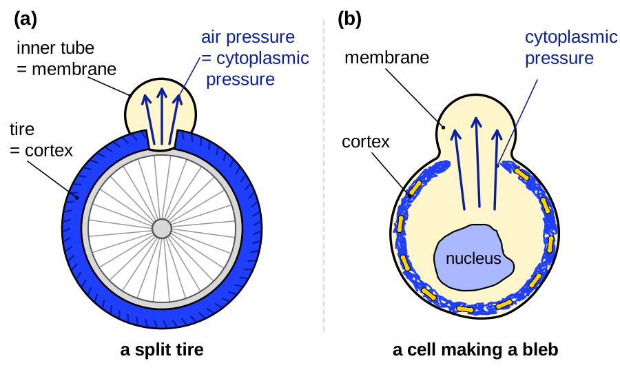
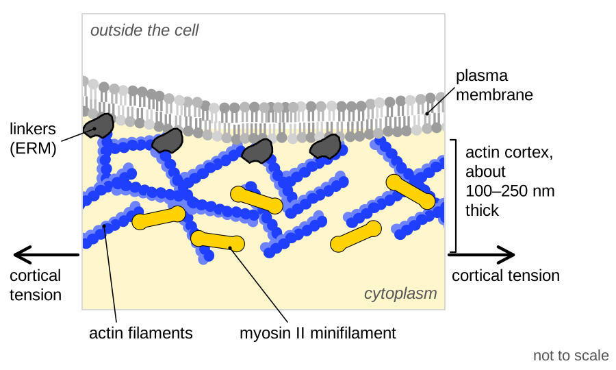
The surface of an animal cell can be pictured as a tire with an inner tube. The plasma membrane, the tube, rests on the cortex, the tire, a thin network of actin filaments and myosin II motors. Myosin pulls on the filaments, so the cortex is under tension, and because it wraps the whole cell this tension pressurizes the cytoplasm (Tinevez et al., 2009). A bleb is an inner tube bulging through a split in the tire: where the cortex fails or the membrane comes unglued from it, pressure pushes out membrane with no actin cortex beneath it (Cunningham, 1995; Keller and Eggli, 1998). That expanding blebs lack an actin cortex is established.
The analogy fails in two ways. The cortex makes its own tension and renews its actin within seconds (Fritzsche et al., 2013), so a cell can mend the split and pull the bleb back. And a lipid membrane ruptures after stretching by about 4% (Sheetz et al., 2006), so a growing bleb gains membrane by peeling it from the cortex (Charras et al., 2008).
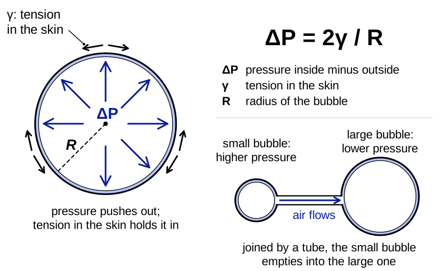
A soap bubble gives a second picture: the tension of its curved skin sets the pressure inside, by Laplace’s law. The cell’s skin, however, is a composite in which the cortex produces the tension, the membrane holds the fluid in and linkers carry the load between them (Brugués et al., 2010).
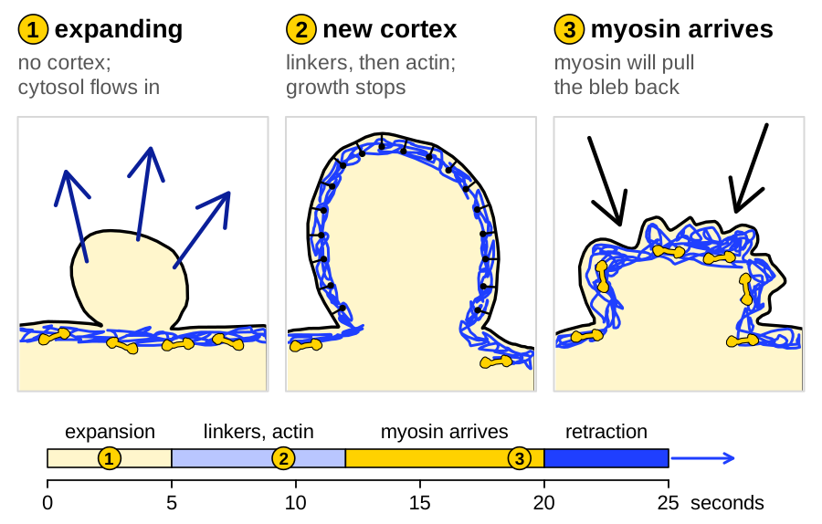
The sections below follow the bleb cycle: the composite fails over a small patch, pressure inflates the bare membrane until growth stops abruptly, and a new cortex pulls the bleb back within about a minute (Cunningham, 1995; Charras et al., 2006). They end with a stable bleb, which holds all these stages at once; measured values are on Blebs by the numbers.
Pressure, tension and the threshold for growth
For a round cell with a thin, uniform cortex, Laplace’s law relates the excess pressure in the cell to cortical tension and cell radius:
where \(P_c\) and \(P_e\) (Pa) are the cytoplasmic and external pressures, \(\gamma_c\) (pN/µm) the cortical tension and \(R\) (µm) the cell radius, assuming an isotropic cortex and one pressure throughout the cytoplasm (Tinevez et al., 2009). By micropipette aspiration, suspended L929 fibroblasts had a tension of 413.6 ± 15.2 pN/µm (mean ± s.e.m., n = 32 cells), implying an excess pressure of about 100 Pa (Tinevez et al., 2009).
Probes that read intracellular pressure directly gave values of the same order: about 400 Pa in blebbing M2 melanoma cells, about 100 Pa after inhibiting Rho-associated kinase, ROCK, with Y-27632 (26 cells each), and 400–600 Pa in HeLa cells (Malboubi et al., 2026).
To find where a bleb can start, Sheetz et al. (2006) treated membrane spanning a circular gap of radius \(a\) (µm) in the cortex as a spherical cap, which at zero bending rigidity inflates when
where \(\sigma_{\mathrm{eff}}\) (pN/µm) is the apparent membrane tension, in-plane tension plus adhesion energy per area. The gap must be wider than the smallest cap the membrane can hold against the pressure, estimated at 0.3–1 µm. The criterion assumes a static cortex and has not been tested directly.
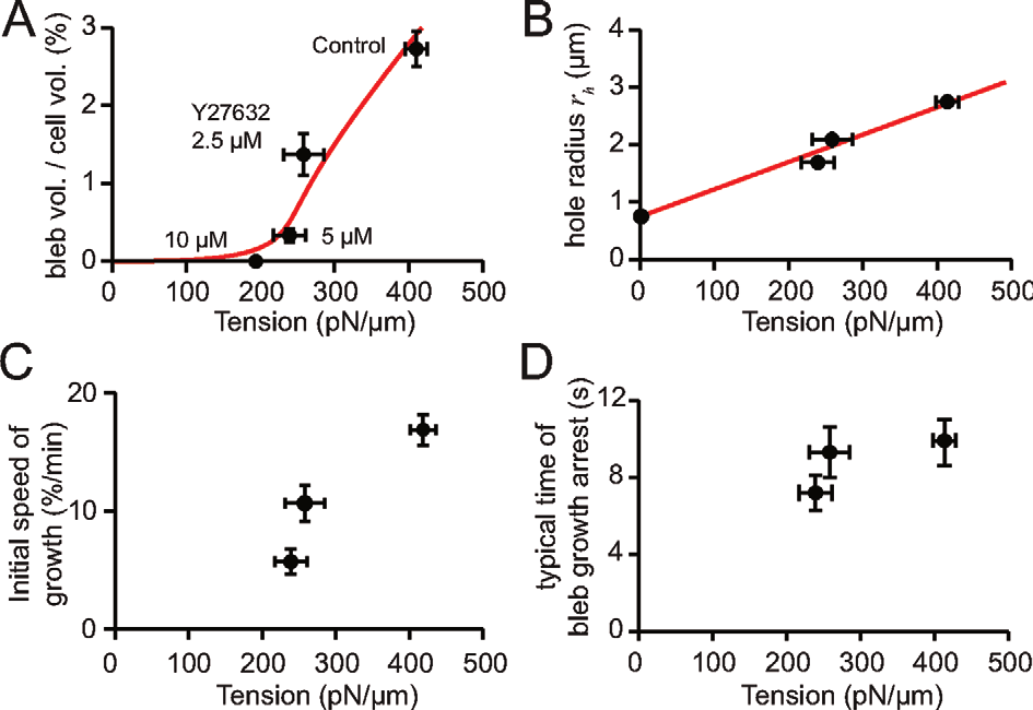
Tinevez et al. (2009) tested whether growth needs a minimum cortical tension by opening a spot about 1.5 µm across in the cortex of suspended L929 cells with a laser. With tension lowered in steps by Y-27632, measured on parallel cells, no bleb grew below about 200 pN/µm, and at 240 pN/µm only about half the cells blebbed. Above that, bleb volume, hole radius and initial growth speed rose with tension, but the time at which growth stopped did not.
The threshold has a simple origin. The membrane over a hole of radius \(r_h\) (µm) can hold at most about \(2\sigma_m/r_h\), so with Eq. 1 growth requires
where \(\sigma_m\) (pN/µm) is the membrane tension, measured by aspiration after the actin drug cytochalasin D. The cortex must be tenser than the membrane by roughly the ratio of cell to hole radius. With \(R \approx 8\) µm, \(r_h \approx 1\) µm and \(\sigma_m \approx 40\) pN/µm this gives about 320 pN/µm, and the full calculation, in which the hole widens with tension, about 200 pN/µm. Eq. 3 combines Eqs. 1 and 2 (our arithmetic), an idea Tinevez et al. (2009) credited to Sheetz et al. (2006).
In the full model, a contractile elastic cortex surrounds a compressible elastic cytoplasm at fixed volume. As the bleb fills, the cell body shrinks and the compressed cytoplasm pushes back, so the pressure available to the bleb falls until it equals what the bleb membrane can hold:
Here \(E_i\) (Pa) and \(\nu_i\) (dimensionless) are the Young’s modulus and Poisson ratio of the cytoplasm, \(E_c\) (Pa) and \(h\) (nm) the modulus and thickness of the cortex, \(\Delta R\) (µm) the decrease in cell radius and \(R_b\) (µm) the bleb radius. Fits gave \(E_c h \approx 240\) pN/µm and \(E_i/(1-2\nu_i) \approx 6{,}750\) Pa (Tinevez et al., 2009).
Eq. 4 is a steady-state balance without cortex turnover or dissipation, so it gives the largest size a bleb could reach. Because the time to arrest, about 7–10 s (read from plot), did not depend on tension, the authors suggested that cortex reassembly stops growth earlier, in which case the fitted moduli are overestimates. A threshold for ablation blebs is strongly supported, but its value rests on one suspended cell type, and metaphase HeLa cells follow the trend without a located threshold (Taneja et al., 2020; Malboubi et al., 2026).
Starting a bleb: detachment and rupture
A bleb can start in two ways: the tube comes unglued from an intact tire, or the tire splits. In cells these are membrane detachment and cortex rupture (Charras and Paluch, 2008), and both have been observed or inferred, in different systems.
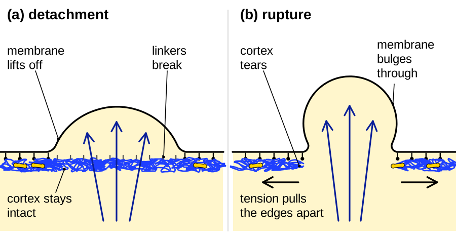
Detachment is supported mainly by images of the cortex left behind. In fixed Walker carcinosarcoma cells, 88.7 ± 7.6% of front blebs kept the old cortex at their base (mean ± s.d.; Keller et al., 2002). Charras et al. (2008) estimated how large a detached patch must be to grow, balancing the work done by pressure against the adhesion lost and the membrane stretched:
Here \(d_c\) and \(\delta_c\) (nm) are the critical diameter and deflection of the patch and \(W_{\mathrm{ad}}\) (J/m²) the membrane–cortex adhesion energy per area; bending is neglected. For \(\Delta P \approx 100\) Pa, \(\sigma_m \approx 2\) pN/µm and a literature value of \(W_{\mathrm{ad}} \approx 6 \times 10^{-6}\) J/m², the patch need only be about 34 nm across and 60 nm deep. Lowering pressure, inhibiting myosin with blebbistatin, raising membrane tension or strengthening attachment each reduced blebbing in M2 cells, as Eq. 5 predicts, although patch size was not measured.
Rupture is supported mainly by perturbation. In nucleus-free L929 fragments, a hole opened in the actin shell before an actin- and myosin-free bulge grew through it (single examples), and local actin drugs raised a bulge on microtubule-depleted L929 cells unless myosin was inhibited (Paluch et al., 2005). In our reading, this shows that a hole suffices, not that natural blebs use one. Walker cells use both routes: with 0.1 µM latrunculin A, 23.5 ± 10.5% of blebs formed where the cortex had first disassembled (mean ± s.d.; Keller et al., 2002).
In Dictyostelium chemotaxing under agarose, two studies disagree. Zatulovskiy et al. (2014) saw the old cortex remain as an intact scar after each bleb formed, whereas Asante-Asamani et al. (2022) found narrow gaps at nucleation sites located from the shape of finished blebs (39 blebs). In our reading, a narrow gap is easy to miss in a survey, but the targeted analysis is correlative and cannot exclude gaps that open just after detachment. Whether migrating vertebrate cells start blebs by rupture is an open question.
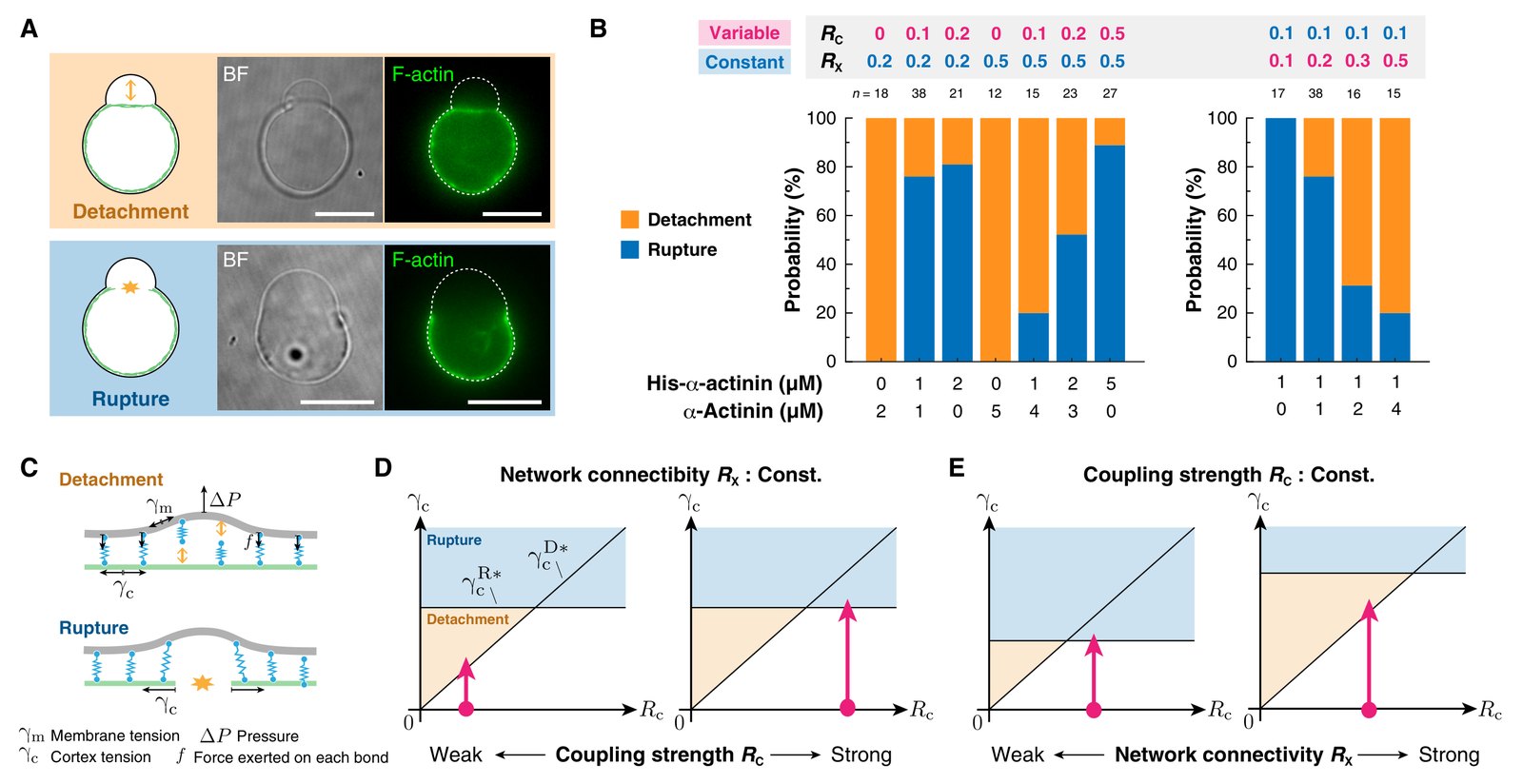
Reconstitution from purified proteins also gives blebs. A contractile cortex tethered to giant vesicles or liposomes blebs more with more myosin and less with stronger linkage (Loiseau et al., 2016; Sakamoto and Murrell, 2024), at tensions estimated 20–400 times below the L929 value (Sakamoto and Murrell, 2024; our arithmetic). Such systems show how one structure can fail either way: in liposomes with a slowly activated cortex, Miyazaki et al. (2026) varied actin–membrane coupling, \(R_C\), and cross-linking, \(R_X\), independently. Rupture rose from 0% to about 89% of first failures as \(R_C\) rose from 0 to 0.5, and fell from 100% to about 20% as \(R_X\) rose from 0.1 to 0.5: gluing the tube more firmly favors a split tire, and a better-knit tire an unglued tube. This switch is strongly supported in vitro, in one study, and untested in cells, and no reconstituted bleb has yet retracted by rebuilding a cortex.
How fast pressure spreads
Whether the pressure drop caused by a growing bleb reaches the whole cell before the bleb stops determines whether blebs act locally or share one pressure. Charras et al. (2005) proposed that cytoplasm behaves like a wet sponge, a poroelastic material in which pressure relaxes only as cytosol flows through the pores of the mesh, so a disturbance spreads diffusively:
Here \(x\) (µm) is the distance reached in time \(t\) (s), \(D\) (µm²/s) the poroelastic diffusion constant, \(k\) (m²/(Pa·s)) the hydraulic permeability, \(K\) (Pa) a bulk modulus of the network and \(\phi\) (dimensionless) the fluid fraction. Relaxation over a length \(L\) takes about \(L^2/D\), assuming linear, isotropic poroelasticity; a stiffer mesh or larger pores, set mainly by actin filaments, speed it (Moeendarbary et al., 2013).
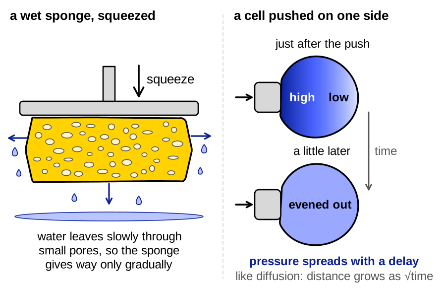
That the cytoplasm is poroelastic is strongly supported by osmotic and indentation tests on HeLa cells, which gave diffusion constants of about 5 and 41 µm²/s (Charras et al., 2009; Moeendarbary et al., 2013). Over 10 µm these give about 20 s and 2.5 s (our arithmetic), comparable to the time a bleb takes to grow, so the material alone cannot say whether a bleb outruns its own pressure drop.
Experiments on blebbing cells point in different directions. Drugs perfused over part of M2 cells reduced blebbing only on the treated side, which Charras et al. (2005) read as pressure not equilibrating over about 10 µm and 10 s. M2 cells beside a pipette that drained pressure also kept blebbing for over 6 min (19 of 19 cells; Malboubi et al., 2026). In contrast, electroporating one site of Entamoeba histolytica stopped blebbing across the cell at once (Maugis et al., 2010), and a second ablation bleb in L929 cells was about 25% smaller wherever it formed, which Tinevez et al. (2009) read as fast equalization. Models, however, reproduce that result both with slow equalization in a poroelastic cytoplasm (Strychalski and Guy, 2016) and without any pressure (Spangler et al., 2011). Whether pressure equalizes faster than blebs grow is an open question.
What stops a bleb growing
A bleb stops growing when a new actin cortex assembles under its membrane (established). Independent laboratories have shown this link directly in three ways. Blocking actin polymerization with cytochalasin D let M2 melanoma blebs grow past the size predicted by their expansion rate (about four blebs; Cunningham, 1995), and acute block of cortex assembly enlarged blebs somewhat (Charras et al., 2008). When new cortex covered only part of a membrane tongue aspirated from Entamoeba histolytica, only the covered part slowed, whereas a fall in pressure or a rise in membrane tension would have acted on the whole tongue (Brugués et al., 2010). And at the edge of cytokineplasts, motile fragments that detach from stable blebs of confined HeLa cells, a rise in cortical actin preceded stalling by about 1.5 s (17 cytokineplasts, one representative experiment; García-Arcos et al., 2024b ◆). The drug experiments rest on few blebs and also disassemble the old cortex (Charras et al., 2008), and the other two are observations rather than perturbations, the Entamoeba one described rather than quantified; their agreement, with each other and with the timing studies below, carries the label.
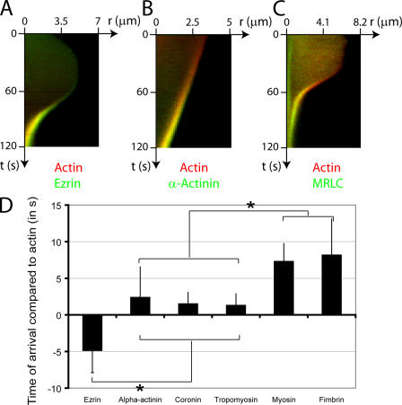
In M2 cells, phalloidin stained blebs that had stopped growing but not growing ones, and Cunningham (1995) concluded that a bleb’s final size reflects a race between inflation and actin polymerization. Ezrin reached the bleb membrane as expansion slowed, and actin followed 4.87 ± 3 s later (s.d., n = 23 blebs; Charras et al., 2006). In Dictyostelium, expanding blebs carried less F-actin than the old cortex (51 blebs) and regained it only once expansion had ceased (Langridge and Kay, 2006).
Brugués et al. (2010) explained why a new cortex can form only once growth slows. They wrote the thickness of a cortex assembling under a moving membrane as
where \(h\) (nm) is the cortex thickness, \(v_p\) (nm/s) the speed at which actin polymerizes at the membrane, \(k_d\) (s⁻¹) the depolymerization rate and \(h_\infty\) (nm) the steady thickness. A meshwork exists only if \(h_\infty\) exceeds the mesh size \(\xi\) (nm). Because depolymerization was assumed to speed up under stress, and a fast membrane stretches the young network, a cortex can form only below a critical membrane speed. Even a thin cortex slows the membrane, which lets more cortex form, so the arrest can be abrupt, as Cunningham (1995) had seen.
A bleb that cannot build a cortex does stop eventually, as actin-free blebs left by a brief pulse of cytochalasin D show (Charras et al., 2006), perhaps through a fall of local pressure (Keller and Eggli, 1998; Charras et al., 2008), slower membrane inflow (Sheetz et al., 2006) or the elastic limits of membrane and cytoplasm found in models without cortex turnover (Strychalski and Guy, 2013; Woolley et al., 2014 abstract only; Strychalski and Guy, 2016). In our reading, these set the larger size a bleb would reach without a cortex.
Rebuilding the cortex and pulling the bleb back
A retracting bleb rebuilds its cortex in a fixed order, as Charras et al. (2006) showed by following 24 cortical proteins on M2 blebs. Ezrin and moesin, two of the ERM proteins that link membrane to actin, arrived first, and the other proteins followed actin within seconds, the myosin regulatory light chain about 7 s after it. The ezrin–actin–myosin order recurred across 401 blebs in vaccinia-infected HeLa cells (Barry et al., 2015) and is strongly supported.
Perturbations separate the roles. Cytochalasin D blocked actin but not ezrin recruitment, an ezrin fragment that blocks ezrin’s binding to actin let the new actin rim tear from the membrane, and under blebbistatin myosin arrived but blebs did not retract (Charras et al., 2006). Ezrin anchors the new cortex, actin builds it and myosin contracts it. Which nucleator builds it is less clear: in ablation-induced blebs, depleting the formin mDia1 halved the speed of new actin assembly, whereas Arp2/3 inhibition doubled it (Bovellan et al., 2014).
Attachment can be measured as an energy. The force \(f_t\) (pN) needed to hold a membrane tether reports an apparent tension,
where \(\kappa\) (J) is the bending rigidity, taken from the literature (2.7–2.9 × 10⁻¹⁹ J); tether force did not depend on tether length between 5 and 10 µm. Tethers from cortex-free membrane give \(\sigma_m\) alone, and the difference gives \(W_{\mathrm{ad}}\), the energy of membrane–cortex attachment per area (Dai and Sheetz, 1999; Diz-Muñoz et al., 2010).
In zebrafish prechordal plate progenitors, \(W_{\mathrm{ad}}\) was about 4.6 × 10⁻⁵ J/m² (our conversion), and loss of ERM function cut it three- to fourfold without changing cortical tension and enlarged blebs in the embryo (Diz-Muñoz et al., 2010). That the balance between attachment and pressure controls bleb formation is strongly supported, although tearing angles in M2 cells gave a lower energy, 1.3–9.8 × 10⁻⁶ J/m² (Charras et al., 2008).
A stable bleb maps the bleb cycle onto space
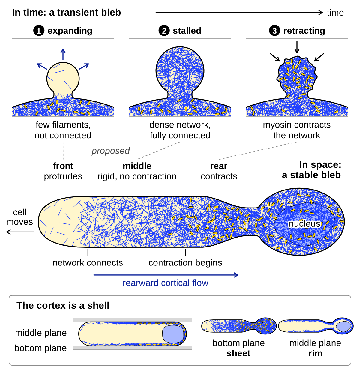
Confined or highly contractile cells can form stable blebs, large elongated blebs that persist for hours and keep protruding although a rearward-flowing cortex lines most of their length (confinement and stable blebs). García-Arcos et al. (2024b ◆) followed this cortex down to single filaments in confined HeLa cells and proposed a mechanism, advected percolation, for how one bleb holds a growing front and a contracting rear.
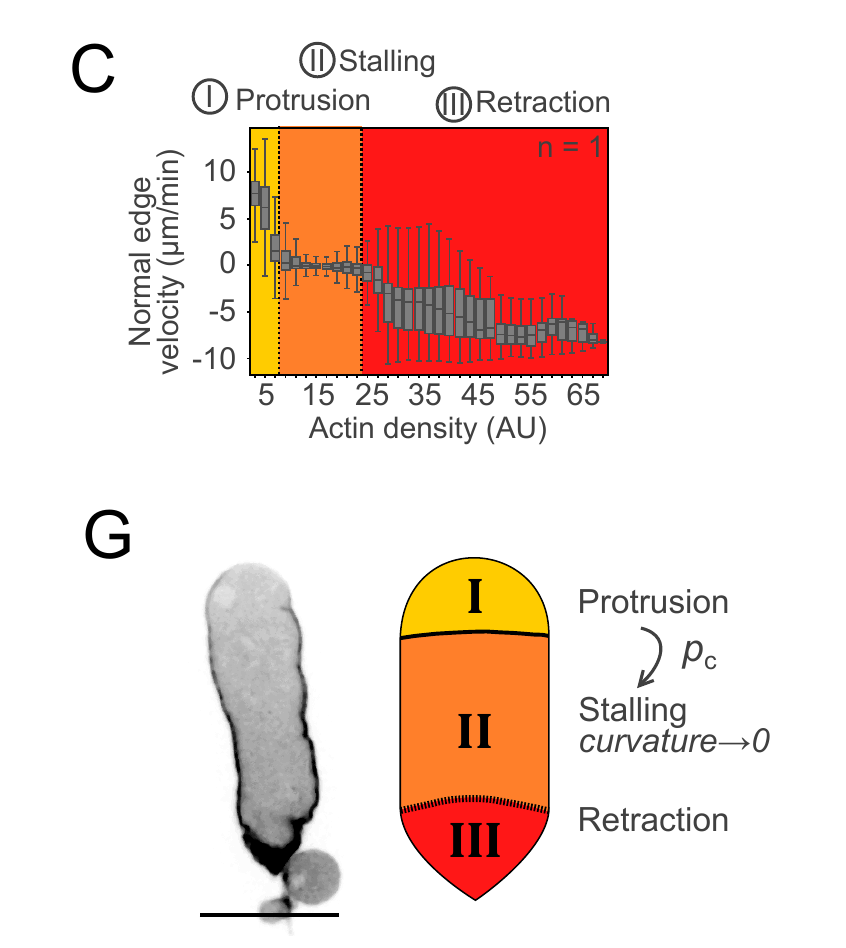
Imaging resolved three zones: at the front, short filaments (0.65 ± 0.45 µm) that diffused and rotated on almost bare membrane until they joined a cluster; in the middle, a network that flowed rearward as a block, without myosin; and at the rear, concentrated myosin IIA and converging flow, as expected where a network contracts. The authors called these the gas-like, solid-like and contractile phases of one cortex.
Percolation, the sudden appearance of a connected path across a system as randomly placed elements become denser, explains how the front becomes the middle. In blebs, the probability of a connection across a measurement window jumped from near 0 to near 1 at an actin occupancy, the fraction of image area showing filament signal, of 0.59, as in simulations of random percolation (91,800 windows, 28 blebs, three experiments). Cluster areas did not fully follow the power law expected at a critical point, so criticality was not claimed.
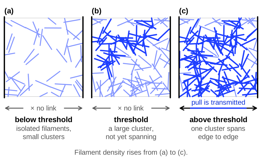
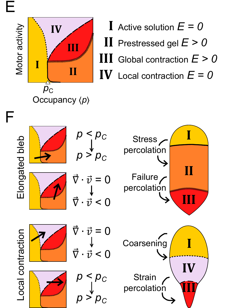
Because it is connected, the middle network carries force from the rear to the front, where filament reorientations and network ruptures, indirect signs of force, were seen. The authors placed the three zones on a state diagram for reconstituted actomyosin gels (Alvarado et al., 2017), in which cross-linking sets connectivity (Alvarado et al., 2013); in the bleb, they proposed, a flow-built gradient of filament density does.
In the advected percolation model, free filaments ahead of the main cluster assemble, diffuse, disassemble and bind rigidly when they meet. The main cluster, all filaments connected to the rear, is a solid carried back at speed \(v_0\) by myosin, and nucleators swept back with it make assembly decay with distance from the rear. In one-dimensional mean-field form (García-Arcos et al., 2024b ◆),
where \(c_a\) is the concentration of free filaments (arbitrary units) at distance \(x\) (µm) from the rear, \(D_a\) (µm²/min) the effective diffusion constant of filaments and nucleators, \(\beta_0\) the assembly rate extrapolated to the rear (concentration per min), \(k_d\) (min⁻¹) the disassembly rate, \(c^{*}\) the percolation concentration (same units as \(c_a\)), \(v_0\) (µm/min) the speed of the rearward flow, \(L_N\) (µm) the decay length of nucleators and \(L_C\) (µm) the forward reach of the connected network. The equation holds ahead of the connected network, where filaments are free. In words, the network reaches as far forward as assembly outpaces disassembly enough for filaments to connect, and faster flow shortens it. The paper writes the logarithm as \(\ln(\beta_0/k_d)\); we include \(c^{*}\) so that its argument has no units.
Comparing \(L_C\) with the bleb length \(L\) (µm) and the length \(L_M\) (µm) over which myosin is concentrated gives the regimes. If \(L_M < L_C < L\), a contractile rear, a rigid middle and a bare front coexist and the bleb is stable; if \(L_C > L\), the rigid network reaches the front and retracts it, as in a transient bleb; and if \(L_C \approx L\) the bleb advances intermittently. Front occupancy sorted these regimes, lying below 0.59 in persistent blebs, near it in intermittent ones and above it in retracting ones (16, 16 and 12 cells). If \(L_C < L_M\), myosin acts on an unconnected network, and slowing nucleation with the Arp2/3 inhibitor CK-666 or the formin inhibitor SMIFH2 produced such local contraction in 42 of 86 and 43 of 97 blebs, against 2 of 27 controls from one experiment.
The model also accounts for how a bleb becomes stable. If actin assembles under a new bleb before myosin is active, the network percolates across the whole bleb and myosin then retracts it; if myosin builds up fast enough, the flow clears the front before it connects, and the bleb stays open. More contraction can therefore stabilize a bleb. In confined HeLa cells, blebs in which rearward flow began before the membrane stalled became stable, and blebs that stalled first, once a new cortex covered their tip, retracted (39 blebs; García-Arcos, 2020 ◆; García-Arcos et al., 2024b ◆, Fig. S7). The percolation threshold and its role in shaping stable blebs are proposed: they rest on one cell line from one laboratory, and no forces were measured, as the authors note.
The bleb cycle laid out in space
García-Arcos et al. (2024b ◆) also proposed that a stable bleb lays out in space the states that a transient bleb passes through in time, relating the three ranges of actin density at which a cytokineplast edge protrudes, stalls and retracts to the gas-like, connected and contractile states of the stable-bleb cortex (Fig. 7C–G). The bare front with its unconnected filaments would correspond to a newly inflated bleb, the connected, prestressed but non-contracting middle to a bleb at the moment growth stops, and the contracting rear to a retracting bleb. The rearward flow converts time into space, because each piece of cortex forms near the front and matures as it travels back. This correspondence is proposed: the states were measured in stable blebs and cytokineplasts, but whether a transient bleb stops at the same occupancy has not been measured.
Blebs pushed from outside
The same physics works with the pressure reversed (our reading). In zebrafish vessel sprouts, the luminal membrane bulged into endothelial cells as inverse blebs, free of F-actin and myosin II until retraction; stopping blood flow stopped the blebbing, and a non-phosphorylatable myosin light chain cut full retraction from about 90% to about 43% of lateral blebs (Gebala et al., 2016). Inverse blebs also form in liver bile canaliculi (Gupta et al., 2017) and between cells of mouse embryos (Schliffka et al., 2024). Pressure-dependent inverse blebs retracted by myosin II are strongly supported; their proposed role as hydraulic pumps rests on bleb position and timing, without measured pressure or flow.
Models: assumptions and tests
None of these models follows a whole cycle with cortex turnover, a poroelastic cytoplasm and parameters from one cell type (our reading).
| Model | What it assumes | What it predicts | Tested? |
|---|---|---|---|
| Laplace instability of a bare membrane (Prothero and Spencer, 1968) | No cortex; membrane tension independent of stretch over a range; constant volume | A bulge grows until tension rises with stretch | Pressure (≈1 Pa) and tension (10 pN/µm) assumed, not measured; later revised into cortex-based models |
| Hole criterion, Eq. 2 (Sheetz et al., 2006) | Static cortex with a circular gap; spherical cap; uniform pressure | Blebs where gaps exceed \(2\sigma_{\mathrm{eff}}/\Delta P\) (estimated 0.3–1 µm) | Not tested directly; equivalent to Eq. 3 (our arithmetic) |
| Elastic cortex and cytoplasm, Eqs. 3–4 (Tinevez et al., 2009) | Contractile cortex without turnover; compressible elastic cytoplasm; fixed volume; uniform pressure | Threshold tension ≈200 pN/µm; bleb volume rising with tension; growth stops at elastic balance | Fits ablation data in one cell type (L929); the tension-independent arrest time lies outside the model, and the authors suggested that cortex reassembly stops growth earlier |
| Detached-patch energy, Eq. 5 (Charras et al., 2008) | Constant pressure and membrane tension at short times; bending neglected | Critical patch ≈34 nm; less blebbing with lower pressure or higher tension or adhesion | Direction confirmed with sucrose, blebbistatin, wheat germ agglutinin and active ezrin (M2 cells); patch size not measured |
| Membrane peeling (Alert and Casademunt, 2016) | Static cortex under a nearly flat membrane; elastic linkers that unbind faster under force, so edge linkers carrying the inflated membrane peel first | Critical radius \(r_p = 2d f^{*}/\Delta P\) (\(d\), bond footprint; \(f^{*}\), pressure at which linkers fail), independent of membrane tension and mostly smaller than the energetic radius | Not tested |
| Poroelastic cytoplasm, Eq. 6 (Charras et al., 2005; Moeendarbary et al., 2013) | Linear, isotropic porous elastic solid soaked in cytosol | Pressure relaxes in a time of order \(L^2/D\); faster with larger pores | Osmotic and indentation tests in HeLa and other cells, without blebs or pressure measured (Charras et al., 2009; Moeendarbary et al., 2013) |
| Viscous cytoplasm and porous cortex (Strychalski and Guy, 2013) | Two-dimensional cell; Newtonian cytoplasm; linear elastic membrane and cortex, the cortex with constant active tension and slipping through the cytosol with drag; linkers removed over a 2 µm patch; no cortex turnover | Growth stops when the elastic membrane balances pressure, at a size set only by membrane and cortex stiffness; formation time set by cortex drag or, at high viscosity, by viscosity; cortex permeability ≈10⁻⁷ µm²; second blebs as large as the first | Formation times of 5–30 s matched by choosing the drag; predicted cortex pores (≈3 nm) far smaller than seen by electron microscopy; no new data |
| Two-dimensional poroelastic cell (Strychalski and Guy, 2016) | Membrane, cortex, linkers and poroelastic cytoplasm; no cytoskeletal turnover | Disturbance crosses the cell in <1 s; equilibration ≈20 s while the bleb reaches 90% of its size at 12.85 s; expansion time ∝ \(1/(Kk)\); smaller second bleb; growth stopped by the elastic response of the cytoplasm | Reinterprets two experiments; no new data |
| Membrane on an elastic network, no pressure (Spangler et al., 2011) | Particle model; vesicles 70–160 nm; no cytosol flow or volume constraint | Blebs above an area mismatch; one bleb favored over two | Qualitative; shows a smaller second bleb need not mean pressure release |
| Cortex regrowth under a moving membrane, Eq. 7 (Brugués et al., 2010) | Cortex thickness set by polymerization at the membrane and stress-accelerated depolymerization; linear viscoelastic cortex; force-dependent linkers | A cortex forms only below a critical membrane speed and then slows the membrane further, so arrest can be abrupt; retraction and self-detachment above two critical thicknesses | Saltatory and oscillating motion of aspirated Entamoeba membrane tongues fitted; partial cortex cover slowed only the covered part; parameters loosely constrained |
| Phase field (Ji et al., 2026) | Coupled membrane and cortex fields; active cortical tension; elastic cytoplasm; fixed volume | Hysteretic onset for detachment blebs, continuous onset when a hole widens with tension; large multiple blebs coarsen into one | Matches L929 ablation volumes from measured hole radii; hysteresis untested |
| Two thresholds (Miyazaki et al., 2026) | Detachment threshold ∝ \(R_C\) (force-dependent bonds); rupture threshold rising with \(R_X\) only (assumed) | Route set by the first threshold that rising tension crosses | Reproduces the in vitro switch; simulated switch at lower \(R_C\) than measured; cells untested |
| Active Kelvin–Voigt cortex recoil (Sakamoto and Murrell, 2024) | After a laser cut, initial recoil speed = active tension / cortex–membrane friction; relaxation time = friction / elasticity | Stronger linkage raises friction and slows recoil | Fits cortex recoil in liposomes, not bleb retraction; 2.2-fold friction rise inferred assuming fixed tension per myosin |
| Advected percolation, Eq. 9 (García-Arcos et al., 2024b ◆) | Filaments diffuse and bind rigidly on contact; connected cluster carried rearward at constant speed; exponential myosin and nucleator profiles; simulated bleb 50 µm long, \(D_a\) = 10 µm²/min, \(k_d\) = 0.5 min⁻¹, \(\beta_0\) not accessible experimentally; no forces computed | Cluster reach \(L_C\) against bleb length and myosin length sets persistent, intermittent or retracting blebs; pulsating front waves; contraction on an unconnected network when nucleation is low | Front occupancy sorted the three regimes (16, 16, 12 cells); CK-666 and SMIFH2 moved the transition rearward; one cell line; forces not measured |
| Inverse-bleb models (Cairoli et al., 2023; Schliffka et al., 2024) | Membrane pocket pressurized from outside; qualitative pseudopotential (embryo) or threshold pressure ∝ cortical tension (vacuoles) | Nucleation as barrier crossing; threshold for giant-vacuole growth | Qualitative only; pressure not measured in the embryo; vacuole model compared qualitatively with published observations |
References (43)
- Alert R, Casademunt J (2016) Bleb nucleation through membrane peeling. Phys Rev Lett 116:068101. doi:10.1103/PhysRevLett.116.068101
- Alvarado J, Sheinman M, Sharma A et al. (2013) Molecular motors robustly drive active gels to a critically connected state. Nat Phys 9:591–597. doi:10.1038/nphys2715
- Alvarado J, Sheinman M, Sharma A et al. (2017) Force percolation of contractile active gels. Soft Matter 13:5624–5644. doi:10.1039/C7SM00834A
- Asante-Asamani E, Grange D, Rawal D et al. (2022) A role for myosin II clusters and membrane energy in cortex rupture for Dictyostelium discoideum. PLoS ONE e0265380. doi:10.1371/journal.pone.0265380
- Barry DJ, Durkin CH, Abella JV et al. (2015) Open source software for quantification of cell migration, protrusions, and fluorescence intensities. J Cell Biol 209:163–180. doi:10.1083/jcb.201501081
- Bovellan M, Romeo Y, Biro M et al. (2014) Cellular control of cortical actin nucleation. Curr Biol 24:1628–1635. doi:10.1016/j.cub.2014.05.069
- Brugués J, Maugis B, Casademunt J et al. (2010) Dynamical organization of the cytoskeletal cortex probed by micropipette aspiration. Proc Natl Acad Sci USA 107:15415–15420. doi:10.1073/pnas.0913669107
- Cairoli A, Spenlehauer A, Overby DR, Lee CF (2023) Model of inverse bleb growth explains giant vacuole dynamics during cell mechanoadaptation. PNAS Nexus 2:pgac304. doi:10.1093/pnasnexus/pgac304
- Charras G, Paluch E (2008) Blebs lead the way: how to migrate without lamellipodia. Nat Rev Mol Cell Biol 9:730–736. doi:10.1038/nrm2453
- Charras GT, Coughlin M, Mitchison TJ et al. (2008) Life and times of a cellular bleb. Biophys J 94:1836–1853. doi:10.1529/biophysj.107.113605
- Charras GT, Hu CK, Coughlin M et al. (2006) Reassembly of contractile actin cortex in cell blebs. J Cell Biol 175:477–490. doi:10.1083/jcb.200602085
- Charras GT, Mitchison TJ, Mahadevan L (2009) Animal cell hydraulics. J Cell Sci 122:3233–3241. doi:10.1242/jcs.049262
- Charras GT, Yarrow JC, Horton MA et al. (2005) Non-equilibration of hydrostatic pressure in blebbing cells. Nature 435:365–369. doi:10.1038/nature03550
- Cunningham CC (1995) Actin polymerization and intracellular solvent flow in cell surface blebbing. J Cell Biol 129:1589–1599. doi:10.1083/jcb.129.6.1589
- Dai J, Sheetz MP (1999) Membrane tether formation from blebbing cells. Biophys J 77:3363–3370. doi:10.1016/S0006-3495(99)77168-7
- Diz-Muñoz A, Krieg M, Bergert M et al. (2010) Control of directed cell migration in vivo by membrane-to-cortex attachment. PLoS Biol 8:e1000544. doi:10.1371/journal.pbio.1000544
- Fritzsche M, Lewalle A, Duke T et al. (2013) Analysis of turnover dynamics of the submembranous actin cortex. Mol Biol Cell 24:757–767. doi:10.1091/mbc.E12-06-0485
- ◆ García-Arcos JM (2020) Stabilization and motility mechanism of blebs in cancer cells. PhD thesis, Université de Paris (Institut Curie). doi:10.70675/a35575c7z85b5z4ad1z98acz50f8d65ebbac
- ◆ García-Arcos JM, Jha A, Waterman CM et al. (2024a) Blebology: principles of bleb-based migration. Trends Cell Biol 34:838–853. doi:10.1016/j.tcb.2024.02.009
- ◆ García-Arcos JM, Ziegler J, Grigolon S et al. (2024b) Rigidity percolation and active advection synergize in the actomyosin cortex to drive amoeboid cell motility. Dev Cell 59:2990–3007.e7. doi:10.1016/j.devcel.2024.06.023
- Gebala V, Collins R, Geudens I et al. (2016) Blood flow drives lumen formation by inverse membrane blebbing during angiogenesis in vivo. Nat Cell Biol 18:443–450. doi:10.1038/ncb3320
- Gupta K, Li Q, Fan JJ et al. (2017) Actomyosin contractility drives bile regurgitation as an early response during obstructive cholestasis. J Hepatol 66:1231–1240. doi:10.1016/j.jhep.2017.01.026
- Ji K, Levine H, Karma A (2026) Phase-field approach to cellular blebbing. Phys Rev E 113:034403. doi:10.1103/gy7s-m4p9
- Keller H, Eggli P (1998) Protrusive activity, cytoplasmic compartmentalization, and restriction rings in locomoting blebbing Walker carcinosarcoma cells are related to detachment of cortical actin from the plasma membrane. Cell Motil Cytoskeleton 41:181–193. doi:10.1002/(SICI)1097-0169(1998)41:2<181::AID-CM8>3.0.CO;2-H
- Keller H, Rentsch P, Hagmann J (2002) Differences in cortical actin structure and dynamics document that different types of blebs are formed by distinct mechanisms. Exp Cell Res 277:161–172. doi:10.1006/excr.2002.5552
- Langridge PD, Kay RR (2006) Blebbing of Dictyostelium cells in response to chemoattractant. Exp Cell Res 312:2009–2017. doi:10.1016/j.yexcr.2006.03.007
- Loiseau E, Schneider JAM, Keber FC et al. (2016) Shape remodeling and blebbing of active cytoskeletal vesicles. Sci Adv 2:e1500465. doi:10.1126/sciadv.1500465
- Malboubi M, Esteki MH, Vaghela MB et al. (2026) The cytoplasm of living cells can sustain transient and steady intracellular pressure gradients. eLife 14:RP105523. doi:10.7554/eLife.105523
- Maugis B, Brugués J, Nassoy P et al. (2010) Dynamic instability of the intracellular pressure drives bleb-based motility. J Cell Sci 123:3884–3892. doi:10.1242/jcs.065672
- Miyazaki M, Laboni FS, Kim T (2026) Reconstitution of actomyosin networks in cell-sized liposomes dissects distinct mechanisms of membrane blebbing and symmetry breaking. Sci Adv 12:eaed8818. doi:10.1126/sciadv.aed8818
- Moeendarbary E, Valon L, Fritzsche M et al. (2013) The cytoplasm of living cells behaves as a poroelastic material. Nat Mater 12:253–261. doi:10.1038/nmat3517
- Paluch E, Piel M, Prost J et al. (2005) Cortical actomyosin breakage triggers shape oscillations in cells and cell fragments. Biophys J 89:724–733. doi:10.1529/biophysj.105.060590
- Prothero JW, Spencer D (1968) A model of blebbing in mitotic tissue culture cells. Biophys J 8:41–51. doi:10.1016/S0006-3495(68)86473-2
- Sakamoto R, Murrell MP (2024) Active tension and membrane friction mediate cortical flows and blebbing in a model actomyosin cortex. Phys Rev Res 6:033024. doi:10.1103/PhysRevResearch.6.033024
- Schliffka MF, Dumortier JG, Pelzer D et al. (2024) Inverse blebs operate as hydraulic pumps during mouse blastocyst formation. Nat Cell Biol 26:1669–1677. doi:10.1038/s41556-024-01501-z
- Sheetz MP, Sable JE, Döbereiner HG (2006) Continuous membrane-cytoskeleton adhesion requires continuous accommodation to lipid and cytoskeleton dynamics. Annu Rev Biophys Biomol Struct 35:417–434. doi:10.1146/annurev.biophys.35.040405.102017
- Spangler EJ, Harvey CW, Revalee JD et al. (2011) Computer simulation of cytoskeleton-induced blebbing in lipid membranes. Phys Rev E 84:051906. doi:10.1103/PhysRevE.84.051906 (read as arXiv:1110.5414v1 preprint)
- Strychalski W, Guy RD (2013) A computational model of bleb formation. Math Med Biol 30:115–130. doi:10.1093/imammb/dqr030
- Strychalski W, Guy RD (2016) Intracellular pressure dynamics in blebbing cells. Biophys J 110:1168–1179. doi:10.1016/j.bpj.2016.01.012
- Taneja N, Bersi MR, Baillargeon SM et al. (2020) Precise tuning of cortical contractility regulates cell shape during cytokinesis. Cell Rep (volume and pages not recorded). doi:10.1016/j.celrep.2020.03.041 (figures not seen)
- Tinevez JY, Schulze U, Salbreux G et al. (2009) Role of cortical tension in bleb growth. Proc Natl Acad Sci USA 106:18581–18586. doi:10.1073/pnas.0903353106
- Woolley TE, Gaffney EA, Oliver JM et al. (2014) Cellular blebs: pressure-driven, axisymmetric, membrane protrusions. Biomech Model Mechanobiol 13:463–476. doi:10.1007/s10237-013-0509-9 abstract only
- Zatulovskiy E, Tyson R, Bretschneider T et al. (2014) Bleb-driven chemotaxis of Dictyostelium cells. J Cell Biol 204:1027–1044. doi:10.1083/jcb.201306147