M192008–2013 · Blebs in migration · established
Blebs framed as a protrusion and migration mode of their own
Between 2008 and 2013, three reviews recast the bleb from a sign of cell death into a pressure-driven protrusion with its own life cycle, and into a migration mode beside the lamellipodium, the flat, actin-pushed protrusion of adherent cells (Charras and Paluch, 2008; Fackler and Grosse, 2008; Paluch and Raz, 2013). They also listed the questions that the field then worked through. That blebs are a migratory protrusion in many cell types is now established (for example, Liu et al., 2015); the reviews’ more specific proposals fared differently.
Before
Until 2008, blebbing was “primarily viewed as a by-product of apoptotic and necrotic processes” (Fackler and Grosse, 2008, p. 879), and blebbing motility had been “eclipsed by its lamellipodial cousin” (Charras and Paluch, 2008, p. 730). The pieces of a different picture existed separately. Bleb structure, stress blebbing, the invasion of rounded tumor cells, local pressure and cortex reassembly had each been studied (M10 to M13), and directed bleb-based migration had just been shown in embryos and amoebae (M18). Most mechanistic knowledge, however, came “from non-migrating cells” (Charras and Paluch, 2008, p. 730). Trinkaus (1973) had seen blebs lead Fundulus deep cells in vivo, and his phrase “Blebs lead the way” (p. 81) later became the title of Charras and Paluch’s review.
What was done
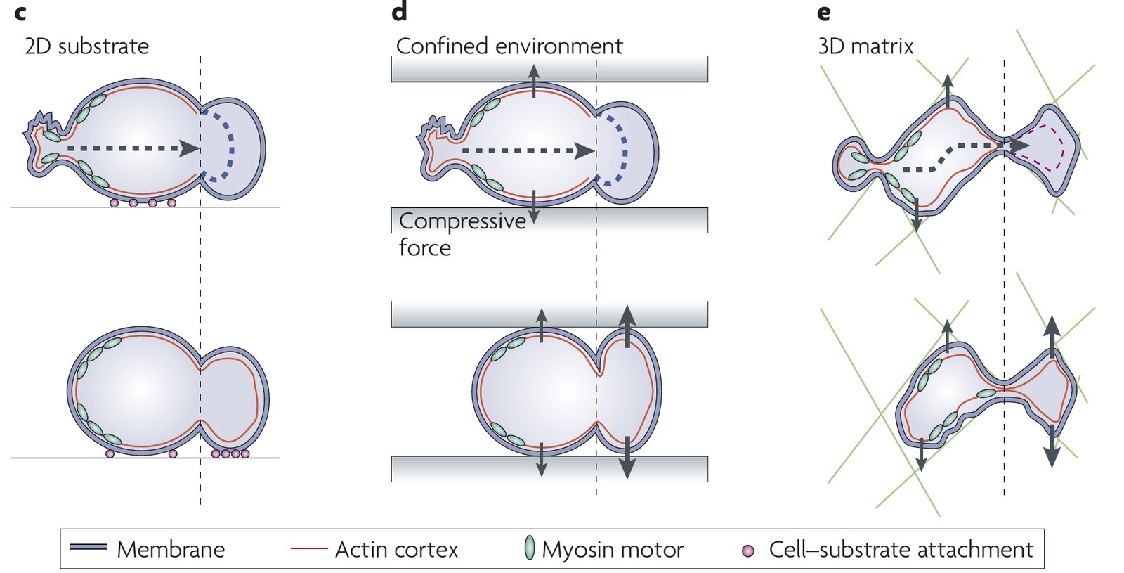
Charras and Paluch (2008), an Opinion article, defined blebs by growth that is “pressure-driven, rather than due to polymerizing actin filaments” (p. 730), cataloged blebbing migrants from embryo cells to leukocytes, and drew a life cycle. A bleb starts by local membrane–cortex detachment or by cortex rupture, expands over 5–30 s, rebuilds a cortex and is retracted by myosin over 60–120 s; in migrating cells, the cell often moves forward instead. Cortex tears had then been seen only in fixed cells. To explain how blebs move a cell, the authors proposed weak adhesion or, in confinement, pushing on the walls without specific adhesion, called chimneying, and they speculated that blebs might be the older protrusion in evolution (p. 734).
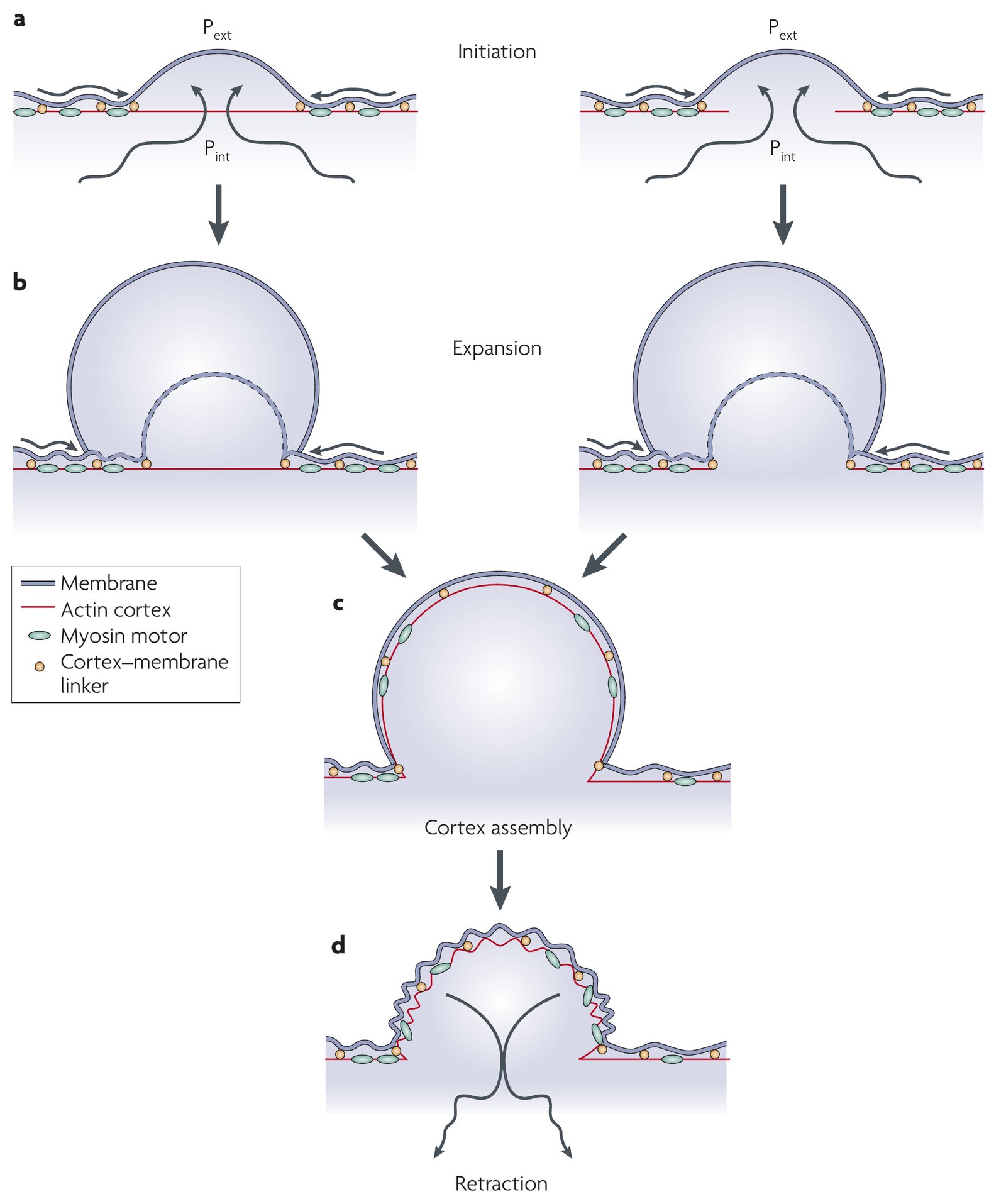
Fackler and Grosse (2008) supplied the molecular side: expansion without actin polymerization, the Rho–ROCK–myosin pathway as core machinery of “virtually all types of blebs” (p. 881), and blebbing as a response to reduced adhesion. Five years later, Paluch and Raz (2013) could be more specific. A bleb, they argued, needs high pressure together with fewer membrane–cortex linkers or breaks in the cortex, its formation is gated by a threshold tension (M12), and cells may use blebs, lamellipodia, both, or switch between them. Of the six proposed ways in which blebs could move a cell, they wrote that “direct experimental evidence for each of these is still scarce” (p. 587).
What it changed
In our reading, the reviews supplied a shared cycle, a vocabulary and a list of questions rather than new mechanisms. When we checked their 45 predictions and open questions against later work, none was unmet or contradicted (table).
| Review | Predictions / questions | Realized | Partly realized | Median delay to realization, years (range) |
|---|---|---|---|---|
| Charras and Paluch, 2008 | 8 / 9 | 8 | 9 | 7 (4–12) |
| Fackler and Grosse, 2008 | 6 / 7 | 5 | 8 | 7 (4–7) |
| Paluch and Raz, 2013 | 7 / 8 | 3 | 12 | 2 (2–7) |
| All three | 21 / 24 | 16 (36%) | 29 (64%) | 7 (2–12) |
This record is flattered, in our reading, by broad questions, by a literature sample partly assembled around the answers, and by answers from the reviewers’ own groups. The clearest success is adhesion-free migration in confinement, realized in vitro (strongly supported). Blebbing Walker cells without focal-adhesion foci moved in high-friction channels but not in low-friction ones (Bergert et al., 2015), and non-adherent neutrophil-like cells pushed about 200–400 Pa into both walls, as chimneying requires (Yip et al., 2015). Yet cells led by a single stable bleb moved mainly by friction between the walls and a rearward-flowing cortex (M20), a route that Paluch and Raz listed and Charras and Paluch had not.
Later work and open questions
Some statements of the reviews were later corrected. Charras and Paluch reported that the actin nucleators Arp2/3 and mDia1 were not detected under blebs of filamin-deficient cells, but isolated bleb cortices from the same cell line held both, and depleting mDia1 enlarged blebs (Bovellan et al., 2014). Adhesion-free migration is not universal either: round breast cancer cells in collagen clustered β1 integrin at 36–58% of bleb necks (Guzman et al., 2020). Two later findings were on no list: the nucleus as a sensor of confinement (Lomakin et al., 2020 ◆; Venturini et al., 2020) and a role for blebs in cell survival (Babiychuk et al., 2011; Weems et al., 2023).
The speculation that blebs are the older protrusion remains an open question. Confined choanoflagellates, the closest living relatives of animals, bleb when Arp2/3 is inhibited (Brunet et al., 2021), but the conservation of the activators of Arp2/3-filled pseudopods across eukaryotes suggests that polymerization-driven crawling is ancient too (Fritz-Laylin et al., 2017). Also still open are which force-transmission mechanism blebbing cells use in tissue, whether blebs are needed for migration and cheaper than lamellipodia, and whether migrating vertebrate cells start blebs by cortex rupture; even in Dictyostelium, two studies disagree on rupture (M15). See also Blebs in cell migration.
References (15)
- Babiychuk EB, Monastyrskaya K, Potez S et al. (2011) Blebbing confers resistance against cell lysis. Cell Death Differ 18:80–89. doi:10.1038/cdd.2010.81
- Bergert M, Erzberger A, Desai RA et al. (2015) Force transmission during adhesion-independent migration. Nat Cell Biol 17:524–529. doi:10.1038/ncb3134
- Bovellan M, Romeo Y, Biro M et al. (2014) Cellular control of cortical actin nucleation. Curr Biol 24:1628–1635. doi:10.1016/j.cub.2014.05.069
- Brunet T, Albert M, Roman W et al. (2021) A flagellate-to-amoeboid switch in the closest living relatives of animals. eLife 10:e61037. doi:10.7554/eLife.61037
- Charras G, Paluch E (2008) Blebs lead the way: how to migrate without lamellipodia. Nat Rev Mol Cell Biol 9:730–736. doi:10.1038/nrm2453
- Fackler OT, Grosse R (2008) Cell motility through plasma membrane blebbing. J Cell Biol 181:879–884. doi:10.1083/jcb.200802081
- Fritz-Laylin LK, Lord SJ, Mullins RD (2017) WASP and SCAR are evolutionarily conserved in actin-filled pseudopod-based motility. J Cell Biol 216:1673–1688. doi:10.1083/jcb.201701074
- Guzman A, Avard RC, Devanny AJ et al. (2020) Delineating the role of membrane blebs in a hybrid mode of cancer cell invasion in three-dimensional environments. J Cell Sci 133:jcs236778. doi:10.1242/jcs.236778
- Liu YJ, Le Berre M, Lautenschlaeger F et al. (2015) Confinement and low adhesion induce fast amoeboid migration of slow mesenchymal cells. Cell 160:659–672. doi:10.1016/j.cell.2015.01.007
- ◆ Lomakin AJ, Cattin CJ, Cuvelier D et al. (2020) The nucleus acts as a ruler tailoring cell responses to spatial constraints. Science 370:eaba2894. doi:10.1126/science.aba2894
- Paluch EK, Raz E (2013) The role and regulation of blebs in cell migration. Curr Opin Cell Biol 25:582–590. doi:10.1016/j.ceb.2013.05.005
- Trinkaus JP (1973) Surface activity and locomotion of Fundulus deep cells during blastula and gastrula stages. Dev Biol 30:68–103. doi:10.1016/0012-1606(73)90049-3
- Venturini V, Pezzano F, Català Castro F et al. (2020) The nucleus measures shape changes for cellular proprioception to control dynamic cell behavior. Science 370:eaba2644. doi:10.1126/science.aba2644
- Weems AD, Welf ES, Driscoll MK et al. (2023) Blebs promote cell survival by assembling oncogenic signalling hubs. Nature 615:517–525. doi:10.1038/s41586-023-05758-6
- Yip AK, Chiam KH, Matsudaira P (2015) Traction stress analysis and modeling reveal that amoeboid migration in confined spaces is accompanied by expansive forces and requires the structural integrity of the membrane-cortex interactions. Integr Biol 7:1196–1211. doi:10.1039/c4ib00245h