M152016–2026 · Blebs as physical objects · strongly supported
Blebs rebuilt from purified proteins, and their two initiation routes separated
Reconstitution asks whether a cortex, the thin network of actin filaments and myosin motors under the plasma membrane, can bleb when it is built from purified proteins inside a liposome, a cell-sized lipid vesicle. Two studies show that a myosin-contracted actin network linked to the membrane is enough (Loiseau et al., 2016; Sakamoto and Murrell, 2024). As in cells (M13), blebbing rises with myosin and falls with stronger membrane–cortex attachment. This is strongly supported, with caveats. A third study separated the two ways a bleb can start (Miyazaki et al., 2026).
Before
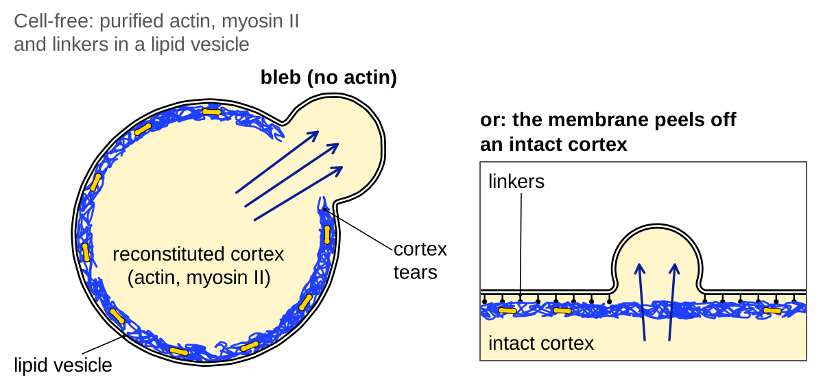
How a single bleb starts was unclear: the membrane might peel away from an intact cortex, or the cortex might tear (rupture versus detachment; Charras and Paluch, 2008). Most front blebs of fixed Walker carcinosarcoma cells kept the old cortex at their base, a sign of detachment (88.7 ± 7.6%, mean ± s.d.; Keller et al., 2002), whereas in nucleus-free L929 fragments a hole opened in the actin shell before a bulge grew through it (single examples; Paluch et al., 2005). What decides between the routes was unknown.
Nor had cell-free systems made a contractile bleb. An actin shell built inside liposomes by the Arp2/3 complex, without myosin, did not deform them (Pontani et al., 2009), and tensed actomyosin shells outside liposomes peeled back or crushed them, with anchoring and cross-linking changing together (Carvalho et al., 2013).
What was done
Loiseau et al. (2016) made the first contractile blebs in vesicles, from actin, muscle myosin II and a fragment of anillin that cross-linked actin and tethered it to a nickel-chelating lipid. Contraction produced one to six small blebs per vesicle, which the authors placed at weakly anchored patches; none formed at high anillin or low myosin. Only end states were imaged.
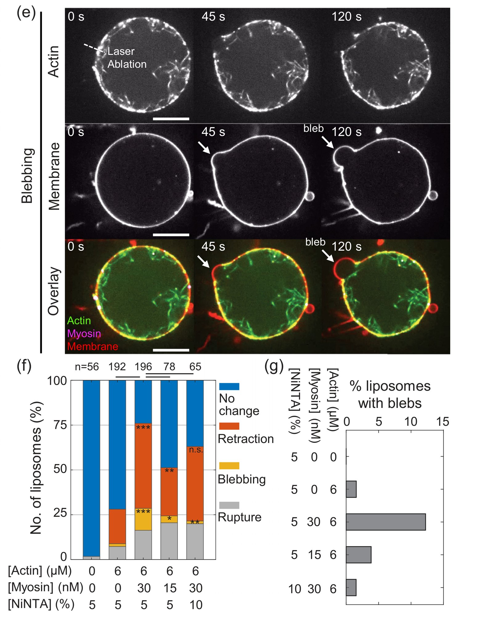
Sakamoto and Murrell (2024) made blebs on demand by cutting a myosin-activated cortex inside liposomes with a laser. Some cuts produced an actin-free bleb, 3.7 ± 2.3 µm across (mean ± s.d., n = 27 blebs). Blebbing rose with myosin and fell when the linking lipid was doubled. Doubling the lipid also slowed recoil of the cut cortex, which the authors, assuming unchanged tension per myosin, read as 2.2-fold higher cortex–membrane friction.
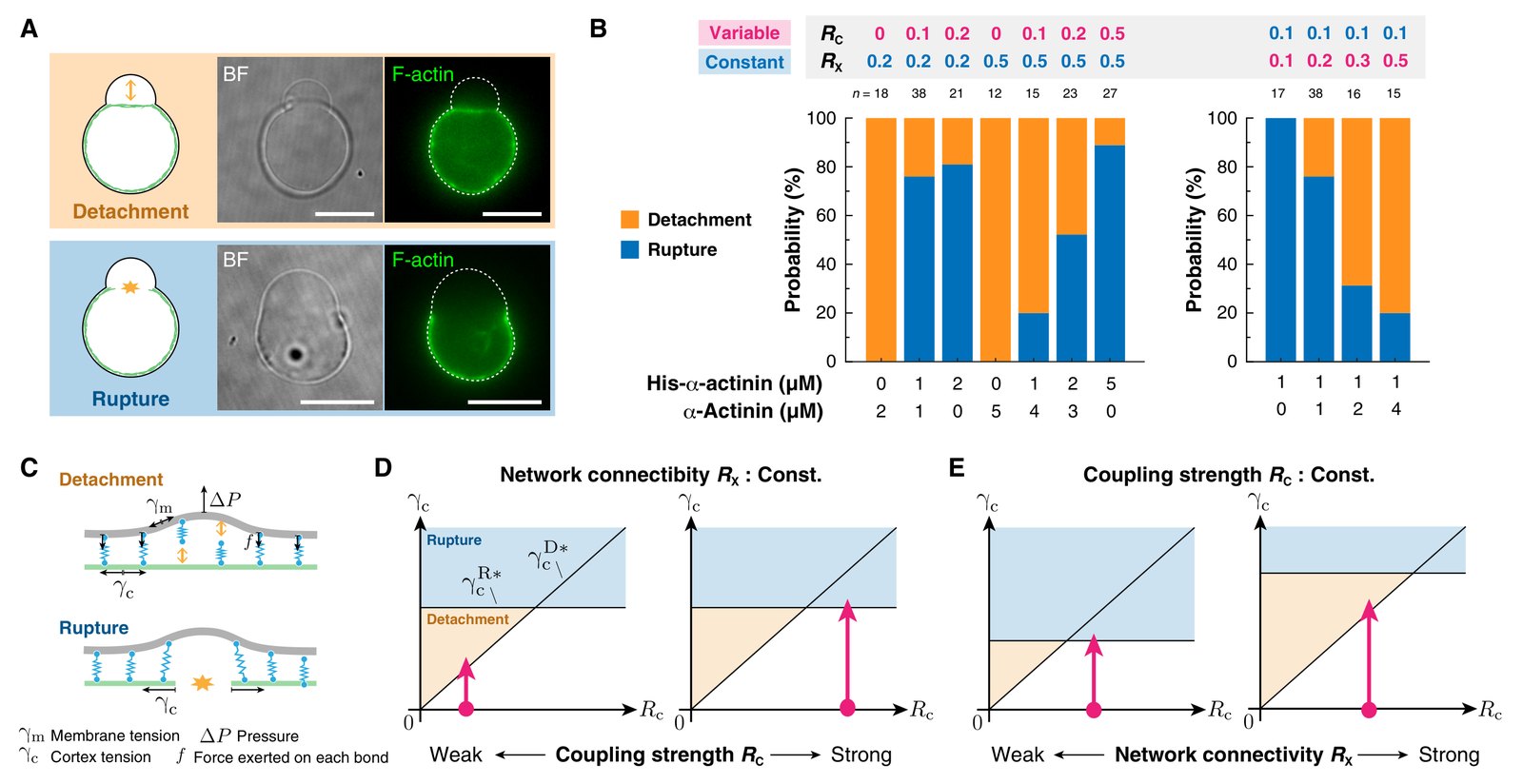
Miyazaki et al. (2026) separated the two factors that the earlier shells had confounded. Two forms of the cross-linker α-actinin, one binding a membrane lipid and one not, set actin–membrane coupling, \(R_C\), independently of cross-linking, \(R_X\). Without coupling, the cortex contracted away and the liposome stayed spherical. With coupling, one composition produced blebs both through tears in the cortex and over locally peeled, intact cortex, and rupture became commoner with more coupling and rarer with more cross-linking.
In the authors’ model, cortical tension pressurizes the liposome (Laplace’s law), and the pressure loads α-actinin bonds that unbind faster under force, so more bonds raise the tension needed to peel the membrane off. The tension needed to tear the cortex was assumed to rise with cross-linking only. Whichever threshold the rising myosin tension crosses first sets the route. An agent-based simulation reproduced the switch, at lower coupling than in experiments.
| Quantity | Value | System | Source |
|---|---|---|---|
| Blebbing after ablation | 1.6%, 4%, 13% at 0, 15, 30 nM myosin (n = 192, 78, 196 per Fig. 1f; labels swapped in Fig. 4d); 12% → 2% at 5% → 10% nickel-chelating (Ni-NTA) lipid (n = 65) | Arp2/3 cortex in liposomes | Sakamoto and Murrell, 2024, Figs. 1f–g, 4d |
| Rupture share of first failures (pixel readout; includes liposomes that never blebbed) | 0%, ≈20%, ≈52%, ≈89% at RC = 0, 0.1, 0.2, 0.5 (RX = 0.5; n = 12, 15, 23, 27); 100%, ≈76%, ≈31%, ≈20% at RX = 0.1, 0.2, 0.3, 0.5 (RC = 0.1; n = 17, 38, 16, 15) | 2D cortex in liposomes | Miyazaki et al., 2026, Fig. 4B |
| Rupture among single blebs | 0/6, 9/18, 19/22 (RC = 0.1, 0.2, 0.5); 6/6, 11/16, 2/10, 0/6 (RX as above) | 2D cortex in liposomes | Miyazaki et al., 2026, fig. S5 |
What it changed
These studies showed that contraction, a membrane–cortex link and a gap in that link suffice to make a bleb. Miyazaki et al. (2026) turned the initiation route, until then put down to cell type, into a matter of two parameters. They also qualified the attachment rule: coupling was needed for any deformation, yet more coupling raised the myosin level at which contraction began. In our reading, links transmit cortical tension to the membrane but also raise the detachment threshold, so coupling acts in both directions (proposed).
Later work and open questions
Reconstituted blebs differ from cellular ones. At best 13% of liposomes blebbed after a cut, and, by our arithmetic, their cortical tensions were 20–400 times below that of L929 cells, whose blebs grow in 10–30 s rather than about 100–170 s (Tinevez et al., 2009; Sakamoto and Murrell, 2024). All three systems used muscle myosins and histidine-tag links rather than the non-muscle myosin II and ERM proteins of cells, and none retracted by rebuilding a cortex. The model’s rupture threshold also rests on severing data that, in our reading, fit a rise of rupture with coupling (Murrell and Gardel, 2012).
In cells, the route appears to depend on the system: besides Walker cells, detachment is supported in MV3 melanoma cells in collagen, where ezrin fell as each bleb appeared (Welf et al., 2020). In Dictyostelium chemotaxing under agarose, two studies disagree. Zatulovskiy et al. (2014) saw no evidence for rupture: the old cortex remained an intact scar for some seconds after each bleb formed. Asante-Asamani et al. (2022) located nucleation sites from the shape of finished blebs and found narrow gaps there, with F-actin near cytoplasmic levels (median relative intensity 1.2, 39 blebs), before or just after detachment. In our reading, a narrow gap is easy to miss in a general survey, whereas the targeted analysis is correlative and cannot exclude gaps that open just after detachment. Whether migrating vertebrate cells start blebs by rupture, and whether cells follow the rule found in liposomes, is an open question (see Physics of blebs).
References (14)
- Asante-Asamani E, Grange D, Rawal D et al. (2022) A role for myosin II clusters and membrane energy in cortex rupture for Dictyostelium discoideum. PLoS ONE e0265380. doi:10.1371/journal.pone.0265380
- Carvalho K, Tsai F-C, Lees E et al. (2013) Cell-sized liposomes reveal how actomyosin cortical tension drives shape change. Proc Natl Acad Sci USA 110:16456–16461. doi:10.1073/pnas.1221524110
- Charras G, Paluch E (2008) Blebs lead the way: how to migrate without lamellipodia. Nat Rev Mol Cell Biol 9:730–736. doi:10.1038/nrm2453
- ◆ García-Arcos JM, Jha A, Waterman CM et al. (2024a) Blebology: principles of bleb-based migration. Trends Cell Biol 34:838–853. doi:10.1016/j.tcb.2024.02.009
- Keller H, Rentsch P, Hagmann J (2002) Differences in cortical actin structure and dynamics document that different types of blebs are formed by distinct mechanisms. Exp Cell Res 277:161–172. doi:10.1006/excr.2002.5552
- Loiseau E, Schneider JAM, Keber FC et al. (2016) Shape remodeling and blebbing of active cytoskeletal vesicles. Sci Adv 2:e1500465. doi:10.1126/sciadv.1500465
- Miyazaki M, Laboni FS, Kim T (2026) Reconstitution of actomyosin networks in cell-sized liposomes dissects distinct mechanisms of membrane blebbing and symmetry breaking. Sci Adv 12:eaed8818. doi:10.1126/sciadv.aed8818 (preprint: doi:10.1101/2025.05.18.654456 preprint)
- Murrell MP, Gardel ML (2012) F-actin buckling coordinates contractility and severing in a biomimetic actomyosin cortex. Proc Natl Acad Sci USA 109:20820–20825. doi:10.1073/pnas.1214753109
- Paluch E, Piel M, Prost J et al. (2005) Cortical actomyosin breakage triggers shape oscillations in cells and cell fragments. Biophys J 89:724–733. doi:10.1529/biophysj.105.060590
- Pontani LL, van der Gucht J, Salbreux G et al. (2009) Reconstitution of an actin cortex inside a liposome. Biophys J 96:192–198. doi:10.1016/j.bpj.2008.09.029
- Sakamoto R, Murrell MP (2024) Active tension and membrane friction mediate cortical flows and blebbing in a model actomyosin cortex. Phys Rev Res 6:033024. doi:10.1103/PhysRevResearch.6.033024
- Tinevez JY, Schulze U, Salbreux G et al. (2009) Role of cortical tension in bleb growth. Proc Natl Acad Sci USA 106:18581–18586. doi:10.1073/pnas.0903353106
- Welf ES, Miles CE, Huh J et al. (2020) Actin-membrane release initiates cell protrusions. Dev Cell 55:723–736.e8. doi:10.1016/j.devcel.2020.11.024
- Zatulovskiy E, Tyson R, Bretschneider T et al. (2014) Bleb-driven chemotaxis of Dictyostelium cells. J Cell Biol 204:1027–1044. doi:10.1083/jcb.201306147