M021835–1872 · Seeing amoeboid movement and naming the bleb, 1755–1919 · established
Amoeboid movement becomes a property of protoplasm
Between Dujardin’s description of sarcode in 1835 and the protoplasm doctrine of the 1860s, the shape-changing movement of the amoeba stopped being the peculiarity of one animalcule and became a property of protoplasm, the living substance of cells, shared by white blood cells and eggs. ‘Amoeboid‘ came to name a behavior rather than a kind of organism (Reynolds, 2008). As history this change is established. Its authors also argued about where the force of a protrusion comes from, and the answer pictured as a hernia is the earliest account we found of the idea behind a bleb.
Before
In the 1830s the cell was pictured as a vesicle “mit Membran, Inhalt und Kern” (with membrane, contents and nucleus), the view that Schultze (1861, p. 8) later set out to replace. Ehrenberg’s memoir of 1830 said only that the amoeba changes shape at will (Ehrenberg, 1830, p. 75), but by 1835 he had proposed a mechanism (Dujardin, 1835, p. 344). One spot of the body slackens, and contraction of the rest drives the contents into it, as viscera are driven into a hernia. This, he wrote, was “das ganze Geheimniss des Formenwechsels” (the whole secret of the change of form; Ehrenberg, 1838, p. 126). The model needs a contractile envelope, and that is where the argument began.
What was done
Dujardin (1835) named the living jelly of these animals sarcode. Seeing no skin on the expansions of amoebae, he rejected the hernia: a protrusion “se forme et s’avance en vertu d’une force inhérente” (forms and advances by an inherent force) and then pulls the rest of the body after it (Dujardin, 1835, pp. 359–360).
Ecker (1849) reached the opposite conclusion by watching the contents move. Granules streamed from the far end of an amoeba into a forming process, so contraction of the body, without any outer skin, drove the contents in; in torn pieces of Hydra, fluid was driven by contraction of the rest “wie in einen Bruchsack” (as into a hernial sac; Ecker, 1849, p. 224).
The same movement was then found in animal cells. Lieberkühn (1854) saw colorless blood corpuscles of frog, fish, dog and human put out and withdraw processes. On a heated stage, Schultze (1865) watched white blood cells crawl among the red ones like amoebae, and in the frog mesentery Cohnheim (1867) watched them bulge through a vein wall and detach over more than 2 h.
These observations made a membrane look unnecessary. Schultze defined the cell as “ein Klümpchen Protoplasma, in dessen Innerem ein Kern liegt” (a lump of protoplasm with a nucleus inside; Schultze, 1861, p. 11), although a clear hyaline outer layer was, in white blood cells, the chief seat of movement (Schultze, 1863). Kühne (1864) then showed that protoplasm behaves like muscle: amoebae rounded up under electrical shocks and recovered, and their responses summed and fatigued. He called amoeba protrusions into which granules streamed so-called ‘Bruchsäcke’ (hernial sacs), and saw large hyaline ‘Blasen’ (blisters) leave the margins of strongly stimulated plasmodia.
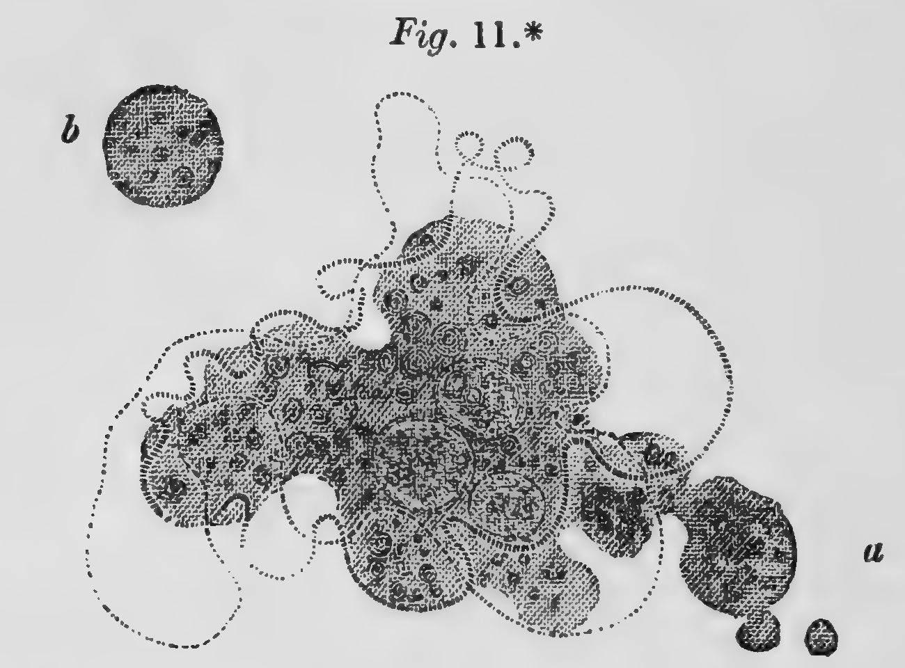
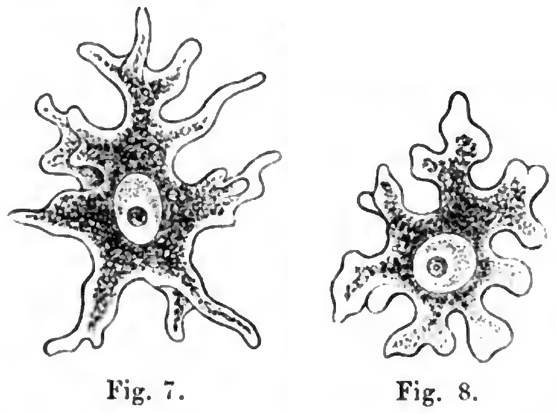
What it changed
The amoeba could now stand for the cell in general (Reynolds, 2008). Carpenter (1865) drew a mucus corpuscle changing shape and shedding a spherule, and Haeckel (1874) judged the creeping egg cell of a sponge indistinguishable from a common amoeba (figures). In pathology, Cohnheim (1867) took pus cells to be white cells that had emigrated mainly by their own contractility, whereas Caton (1870), who saw white cells in inflamed fish move actively yet never emigrate, made the pressure of congestion the deciding force.
The question then became where the contraction happens. Reichert (1862) made pseudopodia semisolid filaments along which contraction travels as a wave. Wallich (1863, pp. 369–370) put it in the outer layer of Amoeba, leaving the interior passive. Heitzmann proposed in 1873 a contracting reticulum that drives fluid against a thin elastic surface layer (Heitzmann, 1891). In our reading, Ehrenberg’s hernia, Ecker’s hernial sac and Kühne’s Bruchsäcke and Blasen describe one sequence, a protrusion pushed out at a weak spot by contraction of the rest of the body (proposed). The gel-tube model of the 1920s (M04) and the pressure models of blebs (M07) later made this sequence quantitative.
Later work and open questions
Several premises did not survive. The membrane returned when Mast (1926) described a plasmalemma separated from the gel beneath it by a fluid layer (M04). Heitzmann’s reticulum was called a fixation artifact (De Bruyn, 1947), although Dellinger (1909) insisted that the mesh he saw in Amoeba was real. Reynolds (2008) credits Wharton Jones (1844) with first comparing amoebae and white corpuscles, but that report mentions no amoebae.
Two questions remain open. Were the hernial sacs, Blasen and leukocyte processes of this period blebs? The texts cannot show whether a membrane separated from an underlying cortex, the criterion used since the late 1990s. And is amoeboid movement one mechanism in amoebae and white blood cells, as De Bruyn (1947) assumed, or a convergent behavior, as Reynolds (2008) argues? We know of no study that tests this (open question). The word ‘bleb’ entered this literature in the 1890s (M03).
References (21)
- Carpenter WB (1865) A Manual of Physiology, including Physiological Anatomy, 4th ed. London: Churchill, pp. 140–150. No DOI.
- Caton R (1870) Contributions to the cell-migration theory. J Anat Physiol 5:35–47. PMID 17230884.
- Cohnheim J (1867) Ueber Entzündung und Eiterung. Virchows Arch 40:1–79. doi:10.1007/bf02968135
- De Bruyn PPH (1947) Theories of amoeboid movement. Q Rev Biol 22:1–24. doi:10.1086/395577
- Dellinger OP (1909) The cilium as a key to the structure of contractile protoplasm. J Morphol 20:171–209. doi:10.1002/jmor.1050200202
- Dujardin F (1835) Recherches sur les organismes inférieurs. Ann Sci Nat Zool (2e sér.) 4:343–377. No DOI.
- Ecker A (1849) Zur Lehre vom Bau und Leben der contractilen Substanz der niedersten Thiere. Z Wiss Zool 1:218–245. No DOI.
- Ehrenberg CG (1830) Beiträge zur Kenntniss der Organisation der Infusorien und ihrer geographischen Verbreitung, besonders in Sibirien. Abh Akad Wiss Berlin 1830 (Phys Abh):1–88, Taf. I–VIII. doi:10.5962/bhl.title.143632
- Ehrenberg CG (1838) Die Infusionsthierchen als vollkommene Organismen. Leipzig: Voss. pp. 125–129. No DOI.
- Haeckel E (1874) Anthropogenie oder Entwickelungsgeschichte des Menschen, 1st ed. Leipzig: Engelmann. pp. 93–114, 384–387. No DOI.
- Heitzmann C (1891) Structure of protoplasm. Int Dent J 12(6):341–353. PMC10109900.
- Kühne W (1864) Untersuchungen über das Protoplasma und die Contractilität. Leipzig: Engelmann. No DOI.
- Lieberkühn N (1854) Ueber die Psorospermien. Arch Anat Physiol Wiss Med 1854:1–24, 349–368. No DOI.
- Mast SO (1926) Structure, movement, locomotion, and stimulation in amoeba. J Morphol 41:347–425. doi:10.1002/jmor.1050410205
- Reichert (1862) On the phenomena of motion in the pseudopodia of the Rhizopoda, and especially on the so-called granular movement and the supposed coalescence of the pseudopodia. Ann Mag Nat Hist (3rd ser.) 10:401–414. doi:10.1080/00222936208681350
- Reynolds A (2008) Amoebae as exemplary cells: the protean nature of an elementary organism. J Hist Biol 41:307–337. doi:10.1007/s10739-007-9142-8
- Schultze M (1861) Ueber Muskelkörperchen und das, was man eine Zelle zu nennen habe. Arch Anat Physiol Wiss Med 1861:1–27. No DOI.
- Schultze M (1863) Das Protoplasma der Rhizopoden und der Pflanzenzellen. Leipzig: Engelmann. No DOI.
- Schultze M (1865) Ein heizbarer Objecttisch und seine Verwendung bei Untersuchungen des Blutes. Arch Mikrosk Anat 1:1–42. No DOI.
- Wallich GC (1863) Further observations on an undescribed indigenous Amœba, with notices on remarkable forms of Actinophrys and Difflugia. Ann Mag Nat Hist (3rd ser.) 11:365–371. doi:10.1080/00222936308681441
- Wharton Jones T (1844) Report on the changes in the blood in inflammation, and on the nature of the healing process. Br Foreign Med Rev 18:255–280. No DOI.