Blebs in cell migration
How a cell can move with blebs, step by step (a front and a rear, front extension, grip, rear), and what embryos, amoebae, tumor cells, leukocytes and confined cells show.
A cell moving through a tissue has to push its front forward, grip its surroundings and bring its rear along. Many cells push their front with lamellipodia, flat sheets driven out by polymerizing actin; others use blebs. The cortex, a thin network of actin filaments and myosin motors under the plasma membrane, contracts and pressurizes the cytoplasm, and wherever the membrane loses its hold on the cortex, cytoplasm flows in and inflates a rounded protrusion (Charras and Paluch, 2008). Long seen mainly as a by-product of dying cells (Fackler and Grosse, 2008), blebs are now established as a migratory protrusion in many cell types (Blaser et al., 2006; Liu et al., 2015; Ruprecht et al., 2015).
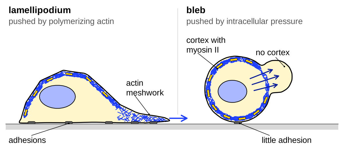
A bleb alone does not move a cell, any more than a lamellipodium does. We therefore follow the four steps that Abercrombie described for crawling fibroblasts, which also apply to bleb-based migration (García-Arcos et al., 2024a ◆): setting up a front and a rear, extending the front, transmitting force to the surroundings and bringing up the rear. We then turn to the systems studied: embryos and amoebae, tumor cells, leukocytes, and confined cells. How a single bleb lives and dies is described in Kinds of blebs.
A front and a rear
To move in one direction, a blebbing cell must bleb more on one side. A bleb starts where the pressurized cytoplasm overcomes the hold of the membrane on the cortex, because the linker proteins let go or the cortex tears. A cell can therefore bias blebbing by loosening membrane–cortex attachment at the front or tightening it at the rear, or by contracting harder near the front (Charras and Paluch, 2008). Both routes have been observed, in different cells.
The clearest evidence concerns the rear. The ERM proteins ezrin, radixin and moesin tie the cortex to the membrane, and in several blebbing cells they gather at the rear and keep blebs away from it. Rounded A375-M2 melanoma cells carried an ezrin-rich, uropod-like structure at the rear, where they blebbed about a third as often per length of outline as elsewhere (4 ± 1.3 against 11 ± 1.4, mean ± s.d. of three experiments), and depleting ezrin reduced invasion (Lorentzen et al., 2011). In mouse T cells that the lipid sphingosine 1-phosphate switches briefly to bleb-based movement, phosphorylated ERM proteins gather at the rear and blebs form at the front, whereas T cells lacking ezrin and moesin bleb all around (Robertson et al., 2021).
How the linkers reach the rear was followed in zebrafish primordial germ cells, which migrate through the embryo with blebs. The front formed first: active Rac and a dense patch of actin, an actin brush, appeared on one side, and the first bleb followed about 50 s later (median, 10 cells). Ezrin reached the opposite side 2–3 min later, apparently carried by the rearward flow of the cortex, and a hyperactive ezrin spread over the whole outline lowered the bleb rate from about 1.7 to 0.7 per minute (read from plot). The authors proposed a feedback loop: the flowing cortex carries bleb inhibitors to the rear, and blebbing at the front sustains the actin polymerization that drives the flow (Olguin-Olguin et al., 2021).
External signals act on this self-organizing system rather than replacing it. In germ cells the guiding chemokine only biased where the front formed (Olguin-Olguin et al., 2021). Calcium was higher at the front, and raising it at the rear produced myosin-dependent blebs there, but that calcium translates the cue into local contraction remains proposed, because a reporter of myosin light-chain phosphorylation differed by only about 7% between front and rear (Blaser et al., 2006).
Where contraction is strongest also differs. In zebrafish germ cells, actin and myosin are enriched at the front, where most blebs start (Grimaldi et al., 2020). In confined cells of many types and in contractile zebrafish progenitors, myosin instead concentrates at the rear (Liu et al., 2015; Ruprecht et al., 2015), and in the progenitors the front holds only a sparse actin network and little ezrin, so it is where the cortex is thinnest and least attached (Ruprecht et al., 2015). A local rise in pressure is therefore not always needed to put blebs at the front.
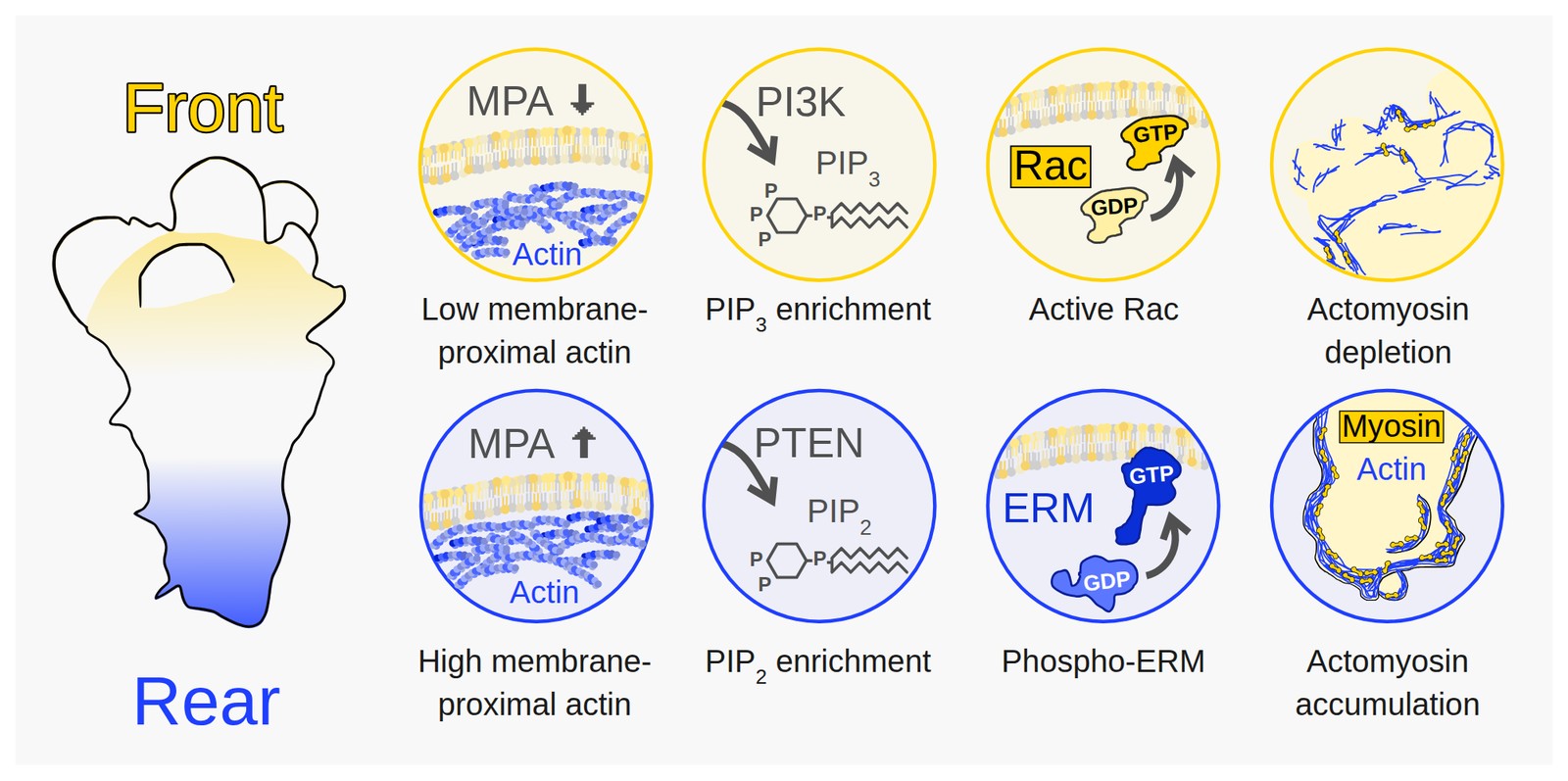
These markers are not laid out the same way in every cell. Inside the leader blebs of confined melanoma cells, active Rac and the lipid PIP3 sit at the base of the bleb and active RhoA and PIP2 at its tip, the reverse of their order in crawling mesenchymal cells (Jha et al., 2026), as described in Blebs as signaling hubs.
Pushing the front forward
In a bleb, the force that pushes the membrane out is the pressure of the cytoplasm, not actin polymerizing against the membrane as in a lamellipodium (Charras and Paluch, 2008). In germ cells inside living zebrafish embryos, a protrusion began where the membrane separated from the actin cortex, cytoplasm carrying small particles flowed into it, and a new cortex then formed under the bleb membrane as the old one faded (Blaser et al., 2006). How cortical tension sets this pressure is explained in Physics of blebs.
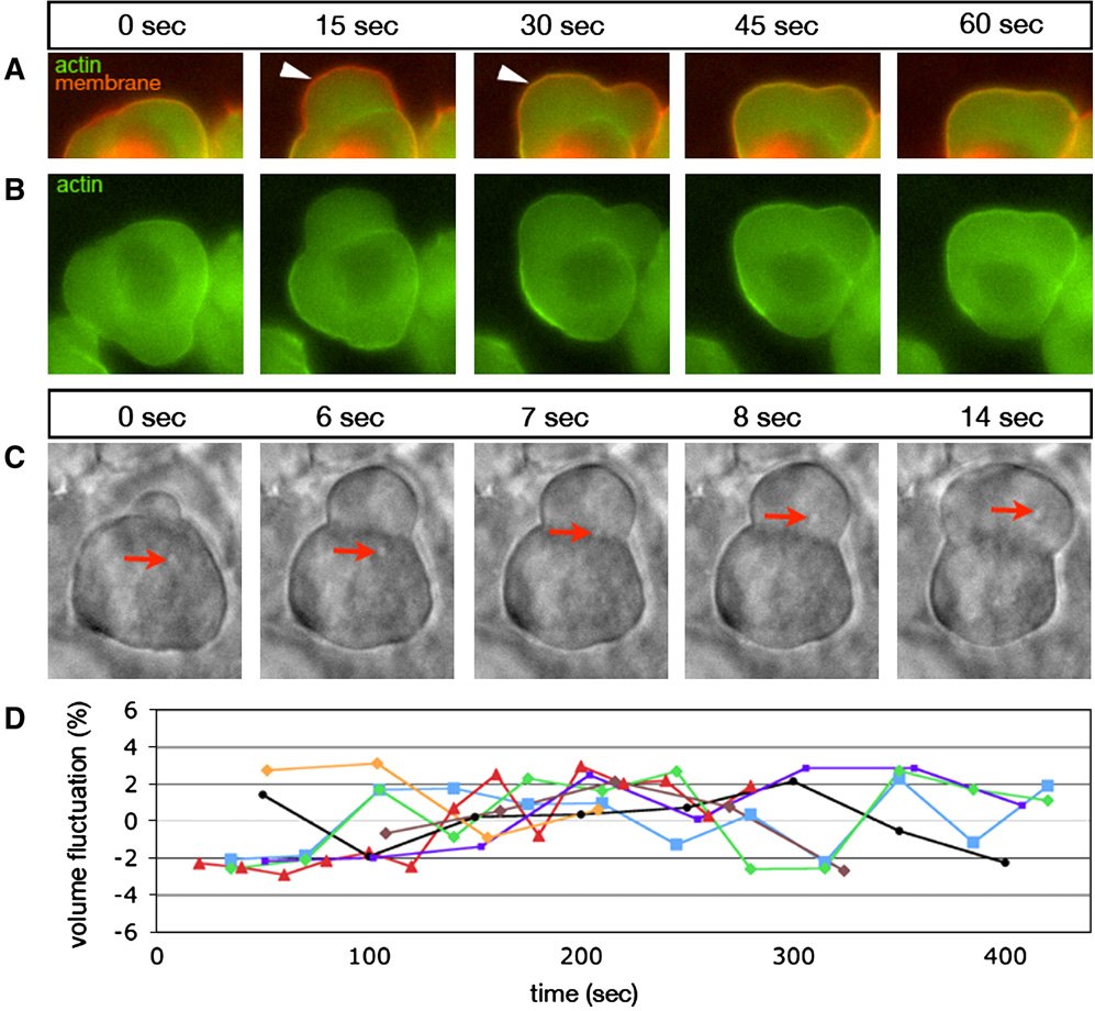
A single bleb, however, advances the front only by its own size, because growth stops once a new cortex forms under its membrane, as seen in Dictyostelium amoebae (Langridge and Kay, 2006); myosin then retracts the bleb, or the cell body moves into it (Charras and Paluch, 2008). Sustained advance therefore needs either a quick succession of blebs or a front where the cortex cannot re-form. On this basis three blebbing regimes have been distinguished: transient blebs, which rebuild a cortex and retract within about a minute, circus blebs, which travel around the cell edge, and stable blebs, whose tip stays free of actin for hours (García-Arcos et al., 2024a ◆). No study has yet compared them systematically.
In stable blebs, myosin at the rear pulls the newly assembled cortex backward, so the tip stays almost bare (Liu et al., 2015; Ruprecht et al., 2015). Imaging single actin filaments in the stable blebs of confined HeLa cells, García-Arcos et al. (2024b ◆) found short, unconnected filaments at the front and, further back, a network carried rearward like a solid. They explained this with percolation: a network transmits the pull of the rear only once enough filaments connect, so the front, below this threshold, keeps protruding. Lowering actin nucleation moved the transition toward the rear. This explanation rests on one cell line.
A growing bleb also needs membrane and a fluid cytoplasm. In zebrafish germ cells, small folds at the front flattened as each bleb inflated, and stabilizing them with a curvature-sensing protein lowered the bleb rate from a median of 1.5 to 0.5 per minute and stopped a median of 29% of germ cells from reaching their target (Goudarzi et al., 2017). In DLD1 cancer cells, calcium entry enlarged expanding blebs, which the authors attributed to a more fluid cytoplasm (Aoki et al., 2021).
Actin polymerization is not excluded from bleb-driven fronts. In melanoma cells in collagen, PI3K and Rac signaling through the Arp2/3 complex enlarged blebs that pushed against the matrix (Driscoll et al., 2024 ◆), and bleb surfaces with the least ezrin advanced fastest. Because ezrin also fell as lamellipodia started, Welf et al. (2020) proposed that releasing membrane–actin links starts protrusions of both kinds, and that pressure or polymerization then provides the push.
Gripping the surroundings
Pushing out blebs does not by itself move a cell. Zebrafish germ cells lacking the protein Dead end neither bleb nor move, and restoring blebbing with myosin light-chain kinase did not restore migration (Goudarzi et al., 2012). Germ cells with weakened E-cadherin function still blebbed and polarized, but hardly moved relative to the tissue (Kardash et al., 2010).
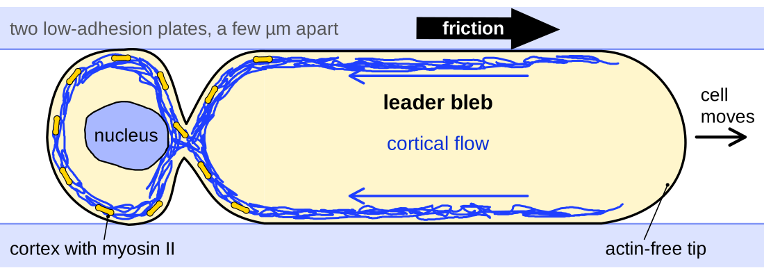
A cell is so small and slow that inertia plays no role, so it moves forward only by pushing backward on its surroundings as hard as they resist its motion (García-Arcos et al., 2024a ◆). In a blebbing cell, the obvious candidate is the cortex flowing from front to rear: coupled to the surroundings, it pushes them backward and the cell forward; slipping, it flows while the cell stays in place. Bergert et al. (2015) tested this with non-adherent, blebbing Walker carcinosarcoma cells in channels 6 × 10 µm in cross-section, whose coatings set the friction. On bovine serum albumin, myosin foci stayed nearly fixed relative to the wall while the cell advanced at 7.6 ± 0.5 µm/min (mean ± s.e.m., n = 18 cells). On Pluronic F127, which lowered the friction coefficient by about four orders of magnitude, the cortex still flowed but the cell did not move. That non-specific friction couples cortical flow to the surroundings is strongly supported in vitro (M20).
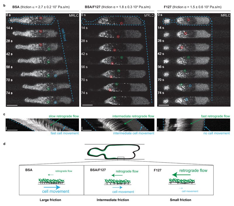
The authors estimated the stress on the wall from a linear friction law, in which the faster the cortex slides past the wall, the larger the stress it transmits:
Here \(\sigma\) (Pa) is the shear stress on the wall, \(\alpha\) (Pa·s/m) the friction coefficient, measured as 2.7 ± 0.2 × 10⁷ Pa·s/m on BSA and 1.5 ± 0.6 × 10³ Pa·s/m on F127 (mean ± s.e.m.), and \(v_{\mathrm{rel}}\) (m/s) the speed of the cortex relative to the wall. The coefficients were measured by pushing unpolarized cells with fluid flow, so they may not hold for migrating cells. The resulting stress, at most about 1 Pa on BSA, is below what traction force microscopy can detect, so it is inferred rather than measured. In the authors’ model, a viscous cortex with a rearward gradient of contractile tension moved the cell only above a threshold friction; it reproduced flows and speeds on all coatings but under-predicted the flow of the surrounding medium (Bergert et al., 2015).
Zebrafish germ cells grip their neighbors more specifically, through E-cadherin. Its knockdown cut the adhesion force between a germ cell and a mesodermal cell from about 17 nN to mostly 1–4 nN (read from plot), whereas germ cells with β1 integrins knocked down still reached their target (Kardash et al., 2010). E-cadherin also brakes the backward flow: without it, actin at the front flowed back faster (about 11 against 6 µm/min, read from plot), blebs formed less focused on the front, and tracks became less persistent, although, unlike in the earlier work, speed did not fall significantly (Grimaldi et al., 2020).
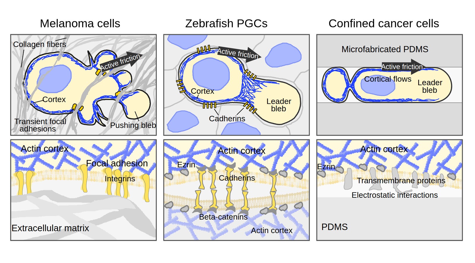
In fibrous matrix, blebbing cells can also use small adhesions. Round breast cancer cells in collagen clustered β1 integrin at about 36–58% of bleb necks (read from plot), and knocking down β1 integrin cut spheroid invasion by 45% (Guzman et al., 2020). Rounded melanoma cells in dense collagen formed small adhesions between blebs, and their large front blebs abraded the collagen without proteases, a process the authors called “worrying” (Driscoll et al., 2024 ◆). Other routes rest on weaker evidence: HL-60 cells squeezed between two non-adhesive gels pushed into both walls, as expected for chimneying, in which a cell braces itself against opposite walls (Yip et al., 2015), and in a two-dimensional model, blebs interlocking with gaps in a discontinuous matrix gained traction without adhesion (Tozluoğlu et al., 2013). Paluch and Raz (2013, p. 587) judged direct evidence for such mechanisms “still scarce”, and in our reading this still holds for cells moving in tissue.
Contraction, disassembly and drag at the rear
A crawling fibroblast must release its rear adhesions before it can follow, but blebbing cells usually have little to release: blebbing Walker cells had no focal adhesion foci and kept their speed after talin knockdown (Bergert et al., 2015). Their rear instead drives the flow: ablating the rear cortex of migrating Walker cells with a laser slowed them by about 80%, and ablating the front by only about 20%, which the authors read as no effect (read from plot; n = 15 and 9; Bergert et al., 2015). Rounded MDA-MB-231 breast cancer cells moving through Matrigel concentrated active myosin in a rear, uropod-like structure whose cortex flowed rearward and dragged matrix beads with it, and inhibiting Rho, ROCK or myosin II strongly reduced invasion. These cells blebbed only at the rear, and the authors considered the blebs a by-product of strong rear contraction (Poincloux et al., 2011).
The flowing cortex must also be dismantled at the rear, or actin would pile up and the flow would stop. In confined cells led by one large bleb, actin assembled at the front and disassembled toward the rear (Liu et al., 2015), and acutely inactivating the actin-severing protein cofilin-1 made actin and myosin pile up at the neck of the leader bleb and slowed the flow (Ullo and Logue, 2021). In melanoma tumors imaged in mice, the actin collar at the base of a front bleb dissolved as the nucleus moved forward, and cells that kept the collar did not translocate (representative cells; Tozluoğlu et al., 2013). The rear can also be a burden: confined cells moved 1.84 times faster after losing a large rear uropod (95% confidence interval 1.32–2.58; Liu et al., 2015).
Embryos and amoebae
Movement led by blebs was described in embryos long before it was analyzed molecularly. Holtfreter (1946) described amphibian embryonic cells whose clear bulges circled around the cell without displacing it, the circus movement. In living Fundulus embryos, leading blebs steered deep cells moving at 2–30 µm/min, and Trinkaus (1973, p. 81) wrote that “Blebs lead the way”.
Zebrafish primordial germ cells, which follow the chemokine SDF-1a to the future gonad (Doitsidou et al., 2002), became the reference system. Raising myosin activation in germ cells gave coiled tracks, and blocking it gave round cells without pseudopods (Blaser et al., 2006). That blebs are a normal, steerable, myosin-driven protrusion of chemotaxing cells is established (M18). The tissue tunes this behavior: in embryos converted to ectoderm, germ cells made fewer blebs and more actin-rich fronts, yet moved as fast (Truszkowski et al., 2023). Germ cells of other animals differ. Mouse germ cells migrated with long actin-rich protrusions (Goodwin et al., 2026), whereas chick germ cells crossed vessel walls with actin-poor, calcium-rich blebs (Morita et al., 2026).
The amoeba Dictyostelium discoideum can use both blebs and actin-filled pseudopods. Its blebbing needs myosin II: no myosin-II-null cell blebbed after a uniform stimulus with cyclic AMP, against about 49% of controls (Langridge and Kay, 2006). Blebs were 20–30% of protrusions in buffer but nearly all under stiff agarose (Zatulovskiy et al., 2014), a switch that ran through Piezo channels and was half-maximal at an applied pressure of about 100 Pa (Srivastava et al., 2020).
Tumor cells
The idea that tumor cells invade with blebs came from a pharmacological dissection. In Matrigel invasion assays, inhibiting Rho-associated kinase (ROCK) almost abolished invasion by rounded, blebbing tumor lines but spared elongated ones; the inhibitor Y-27632 cut invasion by A375m2 melanoma cells to 3.8% of control at depths of 12 µm or more, whereas protease inhibitors did not (Sahai and Marshall, 2003). The authors proposed that contraction raises hydrostatic pressure to push out blebs, but measured neither pressure nor bleb dynamics. That Rho–ROCK contraction drives a rounded, blebbing invasion mode is strongly supported in tumor cell lines in vitro (M11).
This rounded mode is one end of a plastic range. When proteases were blocked, HT-1080 and MDA-MB-231 cells in collagen became spherical and kept migrating, a switch named the mesenchymal–amoeboid transition, although the paper never mentions blebs (Wolf et al., 2003). In A375M2 cells, blocking ROCK made rounded cells elongated without slowing them, and depleting Rac did the reverse (Sanz-Moreno et al., 2008). In our reading, “amoeboid” in this literature means rounded, ROCK-dependent movement with little proteolysis, and whether the cells bleb must be checked paper by paper. Protease independence also depends on the matrix: in cross-linked native collagen, the same kinds of cells needed the protease MT1-MMP (Sabeh et al., 2009).
Whether blebs themselves propel tumor cells is less clear than whether contraction does. Rounded breast cancer cells in Matrigel blebbed only at the rear, whereas in collagen the bleb-neck adhesions and abrading blebs described above took part in invasion, and reducing blebbing with wheat germ agglutinin lowered the fraction of melanoma cells found in tunnels from about 50% to 27% (read from plot; Driscoll et al., 2024 ◆). No perturbation yet removes blebs without also changing contractility or adhesion (Guzman et al., 2020).
Evidence in living animals is indirect. In tumors, ROCK inhibition shifted cells from rounded to elongated movement, but blebs were described rather than measured (Sanz-Moreno et al., 2008). In mammary tumors imaged in mice, ROCK inhibition cut the number of moving cells by about 70%, yet those cells led with an actin-rich front (Wyckoff et al., 2006). In avian embryos, human fibrosarcoma cells crossed the vessel wall with actin-poor blebs or other protrusions, and blocking store-operated calcium entry reduced their extravasation, whereas prostate and breast cancer cells relied on calcium release through IP₃R3 (Morita et al., 2026). Whether blebs as such aid metastasis is an open question.
Leukocytes
Leukocytes were among the first cells proposed to move with blebs, on indirect evidence. In collagen gels, the protrusions of mouse lymphocytes stayed fixed while the cell body passed through a stationary constriction ring in a mean of 30 s (n = 59 cells; Haston et al., 1982), and Haston and Shields (1984), calling the protrusions blebs, proposed that a backward contraction wave drives the cell. This reading is proposed: myosin, pressure and adhesion were not perturbed, and membrane–cortex detachment was not shown (M09). Later, integrin-free dendritic cells, B lymphoblasts and granulocytes crossed collagen at wild-type speed, without imaging of blebs (Lämmermann et al., 2008), and several leukocyte types migrate without myosin II activity, which “largely excludes bleb formation” (Renkawitz and Sixt, 2010, p. 747). In our reading, these cells do not need blebs to move but may use them in particular conditions.
Such conditions are now known. Sphingosine 1-phosphate, the lipid signal for T cells to leave lymphoid organs, raised their intracellular pressure from about 265 to 395 Pa (read from plot) and switched them for a few minutes to bleb-based movement, and T cells lacking ezrin and moesin left lymphoid organs poorly, which the authors attributed to the loss of front-restricted blebbing (Robertson et al., 2021). Neutrophil-like HL-60 cells lacking the branched-actin regulator WAVE advanced by blebs under strong confinement (Graziano et al., 2019). The first candidate in unperturbed tissue is a population of zebrafish epidermal T cells moving in a gap of about 3 µm between keratinocyte layers, with F-actin at the rear and a cytoplasmic bulge at the front; their actin flowed rearward and myosin II or ROCK inhibitors slowed them, but bleb identity was inferred without a membrane or cortex marker (Robertson et al., 2025).
Confinement, stable blebs and leader blebs
Squeezing cells into a narrow space is one of the most reliable ways to make them bleb and move. A non-adherent, blebbing Walker subline migrated only when confined (Bergert et al., 2012). Under a roof 3 µm high on non-adhesive polyethylene glycol, fibroblasts and 20 other cell types, all except normal epithelial cells, produced a fast sub-fraction led by a large bleb that took in most of the cytoplasm. These cells moved at 5.3 ± 1.5 µm/min, against 0.234 ± 0.09 µm/min for mesenchymal cells (mean ± s.e.m.), but only about 10–40% of the cells in each line switched, so confinement makes the switch possible rather than certain (Liu et al., 2015; M20).
Raising contractility produced the same kind of cell. Zebrafish progenitor cells treated with serum or lysophosphatidic acid became pear-shaped “stable-bleb cells” with a cortex-poor front and under agarose moved at 17.1 ± 0.6 µm/min (mean ± s.e.m., n = 67 cells; Ruprecht et al., 2015). Melanoma and other cancer cells confined under agarose formed a single, sausage-shaped leader bleb beyond a thin neck, and knocking down the actin-bundling protein Eps8 lowered cortical tension in rounded cells and, under confinement, shrank the leader bleb from 13.0 ± 0.7% to 7.8 ± 0.6% of the cell-body area (mean ± s.e.m.; n = 144, 168 cells) and about halved the fraction of migrating cells (Logue et al., 2015).
These studies describe the same switch: high contractility and low adhesion produce one large, long-lived bleb that leads the cell, with a cortex flowing from its tip to a contractile rear. The switch is established in vitro; in unperturbed tissue it rests on indirect evidence. In confined HeLa cells, stable blebs can also detach and keep moving as fragments (García-Arcos et al., 2024b ◆), as described in Fragments.
How cells sense confinement seems to differ between cell types. In zebrafish progenitors, the fraction of stable-bleb cells rose from about 23% at 13 µm to 58% at 7 µm (read from plot), and the deformed nucleus was proposed to act as the sensor, activating cytosolic phospholipase A2 and calcium signaling, which raise contractility (Venturini et al., 2020). HeLa cells used the same route, and cytoplasts without a nucleus did not respond (Lomakin et al., 2020 ◆). Neutrophils in narrow channels instead raised RhoA–ROCK–myosin activity at the rear independently of the nucleus, the phospholipase and calcium; blebs were not examined (Deygas et al., 2026). Chick germ cells needed the nuclear route to bleb in the mesentery but not in the vascular plexus (Morimoto et al., 2026 preprint). In our reading, different cells in different tissues use different sensors.
What remains open
Three questions remain. The first is how force is transmitted in tissue: friction, pushing on walls, interlocking with the matrix and swimming have each been shown or modeled in particular settings but never compared in the same three-dimensional environment, so which of them blebbing cells use is an open question (M19). The second is whether blebs are needed for migration or merely accompany high contractility (Paluch and Raz, 2013; Truszkowski et al., 2023). The few perturbations that target blebs rather than contractility, such as stabilizing membrane folds in germ cells or wheat germ agglutinin in melanoma cells, did impair migration (Goudarzi et al., 2017; Driscoll et al., 2024 ◆), but none is entirely specific. The third is whether migration led by stable blebs occurs in unperturbed tissue, where the zebrafish epidermal T cells are the best candidate so far (Robertson et al., 2025).
Whether migrating cells start their blebs by tearing the cortex or by detaching the membrane is discussed in Physics of blebs, the survival signals that blebs carry in Blebs as signaling hubs, and the history of these ideas in A long history of blebbing.
References (51)
- Aoki K, Harada S, Kawaji K et al. (2021) STIM-Orai1 signaling regulates fluidity of cytoplasm during membrane blebbing. Nat Commun 12:480. doi:10.1038/s41467-020-20826-5
- Bergert M, Chandradoss SD, Desai RA et al. (2012) Cell mechanics control rapid transitions between blebs and lamellipodia during migration. Proc Natl Acad Sci USA 109:14434–14439. doi:10.1073/pnas.1207968109
- Bergert M, Erzberger A, Desai RA et al. (2015) Force transmission during adhesion-independent migration. Nat Cell Biol 17:524–529. doi:10.1038/ncb3134
- Blaser H, Reichman-Fried M, Castanon I et al. (2006) Migration of zebrafish primordial germ cells: a role for myosin contraction and cytoplasmic flow. Dev Cell 11:613–627. doi:10.1016/j.devcel.2006.09.023
- Charras G, Paluch E (2008) Blebs lead the way: how to migrate without lamellipodia. Nat Rev Mol Cell Biol 9:730–736. doi:10.1038/nrm2453
- Deygas M, Bernard M, Nivoit P et al. (2026) Adaptive motility enables neutrophils to rapidly navigate confined capillaries. Sci Immunol 11:eaea1426. doi:10.1126/sciimmunol.aea1426
- Doitsidou M, Reichman-Fried M, Stebler J et al. (2002) Guidance of primordial germ cell migration by the chemokine SDF-1. Cell 111:647–659. doi:10.1016/s0092-8674(02)01135-2
- ◆ Driscoll MK, Welf ES, Weems A et al. (2024) Proteolysis-free amoeboid migration of melanoma cells through crowded environments via bleb-driven worrying. Dev Cell 59:2414–2428.e8. doi:10.1016/j.devcel.2024.05.024
- Fackler OT, Grosse R (2008) Cell motility through plasma membrane blebbing. J Cell Biol 181:879–884. doi:10.1083/jcb.200802081
- ◆ García-Arcos JM, Jha A, Waterman CM et al. (2024a) Blebology: principles of bleb-based migration. Trends Cell Biol 34:838–853. doi:10.1016/j.tcb.2024.02.009
- ◆ García-Arcos JM, Ziegler J, Grigolon S et al. (2024b) Rigidity percolation and active advection synergize in the actomyosin cortex to drive amoeboid cell motility. Dev Cell 59:2990–3007.e7. doi:10.1016/j.devcel.2024.06.023
- Goodwin K, Emrich TA, Arnold S et al. (2026) Primordial germ cells experience increasing physical confinement and DNA damage during migration in the mouse embryo. Sci Adv 12:eaec7919. doi:10.1126/sciadv.aec7919
- Goudarzi M, Banisch TU, Mobin MB et al. (2012) Identification and regulation of a molecular module for bleb-based cell motility. Dev Cell 23:210–218. doi:10.1016/j.devcel.2012.05.007
- Goudarzi M, Tarbashevich K, Mildner K et al. (2017) Bleb expansion in migrating cells depends on supply of membrane from cell surface invaginations. Dev Cell 43:577–587.e5. doi:10.1016/j.devcel.2017.10.030
- Graziano BR, Town JP, Sitarska E et al. (2019) Cell confinement reveals a branched-actin independent circuit for neutrophil polarity. PLoS Biol 17:e3000457. doi:10.1371/journal.pbio.3000457
- Grimaldi C, Schumacher I, Boquet-Pujadas A et al. (2020) E-cadherin focuses protrusion formation at the front of migrating cells by impeding actin flow. Nat Commun 11:5397. doi:10.1038/s41467-020-19114-z
- Guzman A, Avard RC, Devanny AJ et al. (2020) Delineating the role of membrane blebs in a hybrid mode of cancer cell invasion in three-dimensional environments. J Cell Sci 133:jcs236778. doi:10.1242/jcs.236778
- Haston WS, Shields JM (1984) Contraction waves in lymphocyte locomotion. J Cell Sci 68:227–241. doi:10.1242/jcs.68.1.227
- Haston WS, Shields JM, Wilkinson PC (1982) Lymphocyte locomotion and attachment on two-dimensional surfaces and in three-dimensional matrices. J Cell Biol 92:747–752. doi:10.1083/jcb.92.3.747
- Holtfreter J (1946) Structure, motility and locomotion in isolated embryonic amphibian cells. J Morphol 79:27–62. doi:10.1002/jmor.1050790103
- Jha A, Chandra A, Farahani PE et al. (2026) CD44 restricts EGFR mobility to polarize cytoskeletal signalling modules driving bleb-based migration. Nat Cell Biol 28:1408–1423. doi:10.1038/s41556-026-01981-1
- Kardash E, Reichman-Fried M, Maître JL et al. (2010) A role for Rho GTPases and cell–cell adhesion in single-cell motility in vivo. Nat Cell Biol 12:47–53. doi:10.1038/ncb2003
- Lämmermann T, Bader BL, Monkley SJ et al. (2008) Rapid leukocyte migration by integrin-independent flowing and squeezing. Nature 453:51–55. doi:10.1038/nature06887
- Langridge PD, Kay RR (2006) Blebbing of Dictyostelium cells in response to chemoattractant. Exp Cell Res 312:2009–2017. doi:10.1016/j.yexcr.2006.03.007
- Liu YJ, Le Berre M, Lautenschlaeger F et al. (2015) Confinement and low adhesion induce fast amoeboid migration of slow mesenchymal cells. Cell 160:659–672. doi:10.1016/j.cell.2015.01.007
- Logue JS, Cartagena-Rivera AX, Baird MA et al. (2015) Erk regulation of actin capping and bundling by Eps8 promotes cortex tension and leader bleb-based migration. eLife 4:e08314. doi:10.7554/eLife.08314
- ◆ Lomakin AJ, Cattin CJ, Cuvelier D et al. (2020) The nucleus acts as a ruler tailoring cell responses to spatial constraints. Science 370:eaba2894. doi:10.1126/science.aba2894
- Lorentzen A, Bamber J, Sadok A et al. (2011) An ezrin-rich, rigid uropod-like structure directs movement of amoeboid blebbing cells. J Cell Sci 124:1256–1267. doi:10.1242/jcs.074849
- Morimoto M, Kamei Y, Morita M et al. (2026) Environment-specific mechanosensing preserves a common bleb-based migratory program across diverse embryonic environments. bioRxiv. doi:10.64898/2026.08.11.744326 preprint
- Morita M, Morimoto M, Ikenouchi J et al. (2026) Bleb-based extravasation uses conserved morphodynamics but divergent calcium control. Nat Commun 17:4422. doi:10.1038/s41467-026-71052-4
- Olguin-Olguin A, Aalto A, Maugis B et al. (2021) Chemokine-biased robust self-organizing polarization of migrating cells in vivo. Proc Natl Acad Sci USA 118:e2018480118. doi:10.1073/pnas.2018480118
- Paluch EK, Raz E (2013) The role and regulation of blebs in cell migration. Curr Opin Cell Biol 25:582–590. doi:10.1016/j.ceb.2013.05.005
- Poincloux R, Collin O, Lizárraga F et al. (2011) Contractility of the cell rear drives invasion of breast tumor cells in 3D Matrigel. Proc Natl Acad Sci USA 108:1943–1948. doi:10.1073/pnas.1010396108
- Renkawitz J, Sixt M (2010) Mechanisms of force generation and force transmission during interstitial leukocyte migration. EMBO Rep 11:744–750. doi:10.1038/embor.2010.147
- Robertson TF, Chengappa P, Gomez Atria D et al. (2021) Lymphocyte egress signal sphingosine-1-phosphate promotes ERM-guided, bleb-based migration. J Cell Biol 220:e202007182. doi:10.1083/jcb.202007182
- Robertson TF, Schrope J, Zwick Z et al. (2025) Live imaging in zebrafish reveals tissue-specific strategies for amoeboid migration. Development 152:dev204351. doi:10.1242/dev.204351
- Ruprecht V, Wieser S, Callan-Jones A et al. (2015) Cortical contractility triggers a stochastic switch to fast amoeboid cell motility. Cell 160:673–685. doi:10.1016/j.cell.2015.01.008
- Sabeh F, Shimizu-Hirota R, Weiss SJ (2009) Protease-dependent versus -independent cancer cell invasion programs: three-dimensional amoeboid movement revisited. J Cell Biol 185:11–19. doi:10.1083/jcb.200807195
- Sahai E, Marshall CJ (2003) Differing modes of tumour cell invasion have distinct requirements for Rho/ROCK signalling and extracellular proteolysis. Nat Cell Biol 5:711–719. doi:10.1038/ncb1019
- Sanz-Moreno V, Gadea G, Ahn J et al. (2008) Rac activation and inactivation control plasticity of tumor cell movement. Cell 135:510–523. doi:10.1016/j.cell.2008.09.043
- Srivastava N, Traynor D, Piel M et al. (2020) Pressure sensing through Piezo channels controls whether cells migrate with blebs or pseudopods. Proc Natl Acad Sci USA 117:2506–2512. doi:10.1073/pnas.1905730117
- Tozluoğlu M, Tournier AL, Jenkins RP et al. (2013) Matrix geometry determines optimal cancer cell migration strategy and modulates response to interventions. Nat Cell Biol 15:751–762. doi:10.1038/ncb2775
- Trinkaus JP (1973) Surface activity and locomotion of Fundulus deep cells during blastula and gastrula stages. Dev Biol 30:68–103. doi:10.1016/0012-1606(73)90049-3
- Truszkowski L, Batur D, Long H et al. (2023) Primordial germ cells adjust their protrusion type while migrating in different tissue contexts in vivo. Development 150:dev200603. doi:10.1242/dev.200603
- Ullo MF, Logue JS (2021) ADF and cofilin-1 collaborate to promote cortical actin flow and the leader bleb-based migration of confined cells. eLife 10:e67856. doi:10.7554/eLife.67856
- Venturini V, Pezzano F, Català Castro F et al. (2020) The nucleus measures shape changes for cellular proprioception to control dynamic cell behavior. Science 370:eaba2644. doi:10.1126/science.aba2644
- Welf ES, Miles CE, Huh J et al. (2020) Actin-membrane release initiates cell protrusions. Dev Cell 55:723–736. doi:10.1016/j.devcel.2020.11.024
- Wolf K, Mazo I, Leung H et al. (2003) Compensation mechanism in tumor cell migration: mesenchymal–amoeboid transition after blocking of pericellular proteolysis. J Cell Biol 160:267–277. doi:10.1083/jcb.200209006
- Wyckoff JB, Pinner SE, Gschmeissner S et al. (2006) ROCK- and myosin-dependent matrix deformation enables protease-independent tumor-cell invasion in vivo. Curr Biol 16:1515–1523. doi:10.1016/j.cub.2006.05.065
- Yip AK, Chiam KH, Matsudaira P (2015) Traction stress analysis and modeling reveal that amoeboid migration in confined spaces is accompanied by expansive forces and requires the structural integrity of the membrane-cortex interactions. Integr Biol 7:1196–1211. doi:10.1039/c4ib00245h
- Zatulovskiy E, Tyson R, Bretschneider T et al. (2014) Bleb-driven chemotaxis of Dictyostelium cells. J Cell Biol 204:1027–1044. doi:10.1083/jcb.201306147