M182006 · Blebs in migration · established
Blebs drive directed migration in embryos and amoebae
In 2006, three studies showed that blebs, driven by myosin II contraction rather than by actin polymerization, are a normal and steerable protrusion of cells that move up a chemical gradient, a behavior called chemotaxis: zebrafish primordial germ cells (PGCs) in the embryo (Blaser et al., 2006) and Dictyostelium amoebae (Langridge and Kay, 2006; Yoshida and Soldati, 2006). That blebs serve as such a protrusion is established; how the chemical cue decides where blebs form is less clear.
Before
At the time, protrusion was explained mainly by actin polymerization at the cell front, and blebs were read mainly as signs of cell death (Charras and Paluch, 2008). Yet blebs had been seen leading embryo cells long before. Charras and Paluch credit this to Holtfreter (1943), which describes neither blebs nor circus movement; it was in 1946 that Holtfreter described amphibian embryonic cells whose clear bulges traveled around the cell without moving it, now called circus movement, and worm-like cells that crept forward by waves of constriction when contact gave them “a sufficient frictional support” (Holtfreter, 1946, p. 43). He rejected internal pressure as the cause.
Trinkaus (1973) then saw, in living Fundulus embryos, deep cells whose blebs, which he proposed to be pressure herniations, led and steered the cells; “Blebs lead the way”, he wrote (p. 81). Blebbing deep cells later moved directly toward an epithelial wound up to 800 µm away, so they could follow a cue (Fink and Trinkaus, 1988). Zebrafish PGCs were known to follow the chemokine SDF-1a, but their protrusions had never been called blebs (Doitsidou et al., 2002; Reichman-Fried et al., 2004).
What was done
In living zebrafish embryos, a PGC protrusion began when the membrane separated from the actin cortex, with no actin at the front. A new cortex then formed beneath the membrane while the old one disassembled, and cytoplasm flowed into the protrusion while the cell’s volume stayed nearly constant. These are the steps of a bleb, pushed by pressure rather than by polymerization (Blaser et al., 2006).
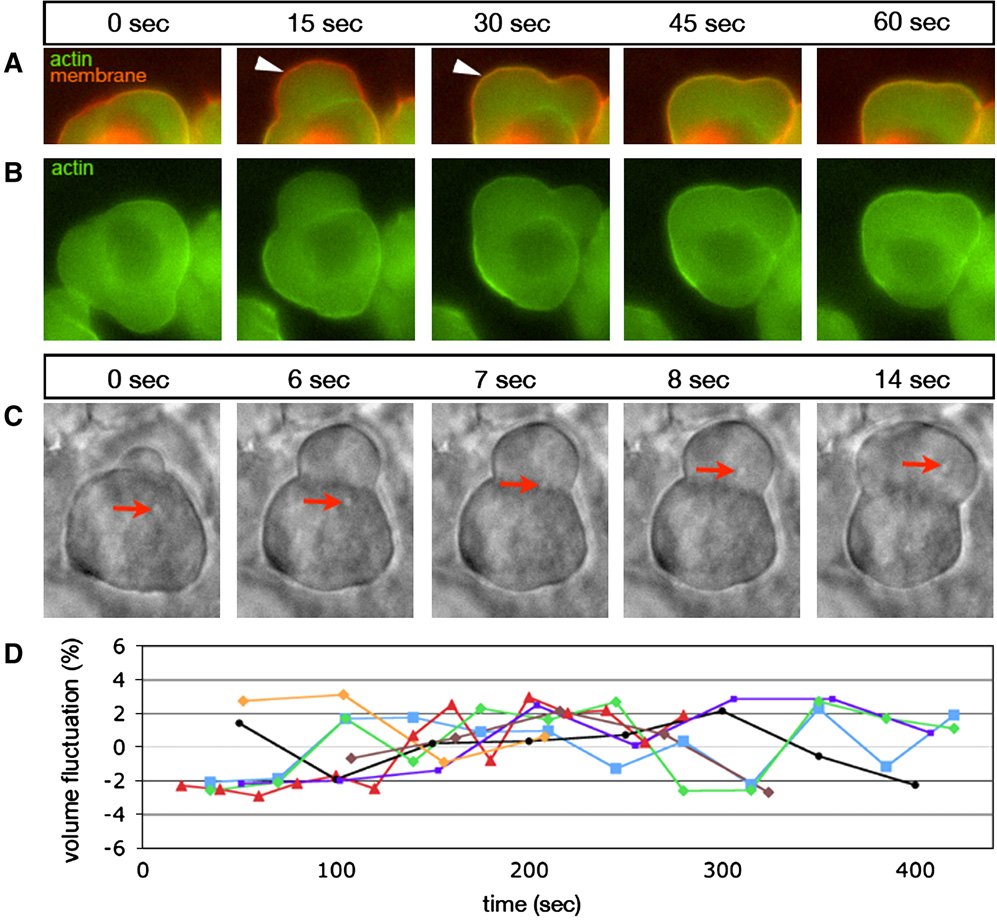
What drives these blebs, and where do they form? Raising myosin activation in germ cells alone gave coiled tracks, whereas dominant-negative kinases or blebbistatin, a myosin II inhibitor, gave round cells without pseudopods. Phosphorylated myosin light chain and free calcium were higher at the front. Calcium fell when SDF-1a or its receptor was knocked down, and raising calcium at the rear produced myosin-dependent blebs there. The authors proposed a chain: the chemokine raises calcium at the front, calcium activates myosin, and contraction produces blebs where the cell is heading.
Two Dictyostelium studies reached a similar conclusion. Cells in low-osmolarity buffer made hemispherical protrusions free of F-actin and blebbed at the front during chemotaxis, whereas myosin-II-null cells, or cells in hyperosmotic sorbitol, made filopodia and lamellipodia (Yoshida and Soldati, 2006). Uniform cAMP, the chemoattractant, made cells round up and bleb without adding surface area or volume, and myosin-II-null cells did not bleb at all (Langridge and Kay, 2006).
| Quantity | Value | System | Source |
|---|---|---|---|
| Relative Ca²⁺ in protrusions, wild type vs SDF-1a morphant | ≈1.47 vs ≈0.93 (a.u.; s.e.m.; n = 16, 18; read from plot) | zebrafish PGCs, 9–12 h post-fertilization | Blaser et al., 2006, Fig. 5F |
| Bleb frequency with calcium entry at the rear, without vs with blebbistatin | ≈3.1 vs ≈0.25 blebs/min (s.e.m.; n = 8, 8; read from plot) | zebrafish PGCs in vivo | Blaser et al., 2006, Fig. 7E |
| Area-gain rate, 0 → 150 mM sorbitol | wild type 1.32 → 0.62 µm²/s; myosin-II-null 0.78 → 0.55 µm²/s (s.e.m.; read from plot) | vegetative Dictyostelium | Yoshida and Soldati, 2006, Fig. 6A |
| Cells blebbing, myosin-II-null vs control | 0% vs ≈49% (P < 0.001; 8 trials, 218 cells) | Dictyostelium | Langridge and Kay, 2006, Fig. 4D |
What it changed
Ridley (2011) named Blaser et al. (2006) the first in vivo demonstration of directed bleb-based migration. Fink and Trinkaus (1988) had come earlier; what Blaser et al. added was the chemokine cue, the bleb cycle and its control by myosin and calcium. Charras and Paluch (2008) then built the three papers into a migration mode of its own (M19).
Later work filled in missing steps. In PGCs, the traction that turns blebs into movement was traced to actin brushes coupled to neighboring cells through E-cadherin rather than integrins (Kardash et al., 2010). Store-operated calcium entry promoted blebbing in cancer cells and chick PGCs (Aoki et al., 2021; Morita et al., 2023 preprint), though some cancer cells used calcium from internal stores (Morita et al., 2026), and Aoki et al. (2021) attributed the larger blebs of colorectal cancer cells to a more fluid cytoplasm, not to stronger contraction. That store-operated calcium entry promotes blebbing is strongly supported in vitro, by one network of laboratories.
Later work and open questions
Whether PGCs keep their volume while blebbing is debated. Taloni et al. (2015) measured volume fluctuations of up to ±10%, damped by inhibiting contraction or water channels, whereas Goudarzi et al. (2019) saw no volume change at bleb events in 15 PGCs and no effect of knocking down water channels. Both rest on few cells, as did the original result from seven slowly sampled cells (Blaser et al., 2006). In our reading, differences in sampling and in what was measured, global fluctuations or single bleb events, may explain the conflict.
How the chemokine positions the front is also less direct than first thought. Active Rac and an actin brush precede the first bleb, and the chemokine biases where this front forms without changing the order of events (Olguin-Olguin et al., 2021). That chemokine-induced calcium defines the front therefore remains proposed.
Finally, blebbing alone does not move a cell: restoring blebbing did not restore movement in PGCs lacking the protein Dead end (Goudarzi et al., 2012). The Dictyostelium tests used drugs that act beyond blebbing and a uniform cAMP shock, not a gradient. Whether blebs are needed for directed migration, or are a side effect of high contractility, is still asked (Paluch and Raz, 2013; Truszkowski et al., 2023). See also Blebs in cell migration.
References (21)
- Aoki K, Harada S, Kawaji K et al. (2021) STIM-Orai1 signaling regulates fluidity of cytoplasm during membrane blebbing. Nat Commun 12:480. doi:10.1038/s41467-020-20826-5
- Blaser H, Reichman-Fried M, Castanon I et al. (2006) Migration of zebrafish primordial germ cells: a role for myosin contraction and cytoplasmic flow. Dev Cell 11:613–627. doi:10.1016/j.devcel.2006.09.023
- Charras G, Paluch E (2008) Blebs lead the way: how to migrate without lamellipodia. Nat Rev Mol Cell Biol 9:730–736. doi:10.1038/nrm2453
- Doitsidou M, Reichman-Fried M, Stebler J et al. (2002) Guidance of primordial germ cell migration by the chemokine SDF-1. Cell 111:647–659. doi:10.1016/s0092-8674(02)01135-2
- Fink RD, Trinkaus JP (1988) Fundulus deep cells: directional migration in response to epithelial wounding. Dev Biol 129:179–190. doi:10.1016/0012-1606(88)90172-8
- Goudarzi M, Banisch TU, Mobin MB et al. (2012) Identification and regulation of a molecular module for bleb-based cell motility. Dev Cell 23:210–218. doi:10.1016/j.devcel.2012.05.007
- Goudarzi M, Boquet-Pujadas A, Olivo-Marin JC et al. (2019) Fluid dynamics during bleb formation in migrating cells in vivo. PLoS One 14:e0212699. doi:10.1371/journal.pone.0212699
- Holtfreter J (1943) Properties and functions of the surface coat in amphibian embryos. J Exp Zool 93:251–323. doi:10.1002/jez.1400930205
- Holtfreter J (1946) Structure, motility and locomotion in isolated embryonic amphibian cells. J Morphol 79:27–62. doi:10.1002/jmor.1050790103
- Kardash E, Reichman-Fried M, Maître JL et al. (2010) A role for Rho GTPases and cell–cell adhesion in single-cell motility in vivo. Nat Cell Biol 12:47–53. doi:10.1038/ncb2003
- Langridge PD, Kay RR (2006) Blebbing of Dictyostelium cells in response to chemoattractant. Exp Cell Res 312:2009–2017. doi:10.1016/j.yexcr.2006.03.007
- Morita M, Morimoto M, Teramoto T et al. (2023) The SOCE system is critical for membrane bleb formation to drive avian primordial germ cell migration. bioRxiv. doi:10.1101/2023.06.12.544577 preprint
- Morita M, Morimoto M, Ikenouchi J et al. (2026) Bleb-based extravasation uses conserved morphodynamics but divergent calcium control. Nat Commun 17:4422. doi:10.1038/s41467-026-71052-4
- Olguin-Olguin A, Aalto A, Maugis B et al. (2021) Chemokine-biased robust self-organizing polarization of migrating cells in vivo. Proc Natl Acad Sci USA 118:e2018480118. doi:10.1073/pnas.2018480118
- Paluch EK, Raz E (2013) The role and regulation of blebs in cell migration. Curr Opin Cell Biol 25:582–590. doi:10.1016/j.ceb.2013.05.005
- Reichman-Fried M, Minina S, Raz E (2004) Autonomous modes of behavior in primordial germ cell migration. Dev Cell 6:589–596. doi:10.1016/s1534-5807(04)00074-7
- Ridley A (2011) Blebs on the move. Dev Cell 20:e1. doi:10.1016/j.devcel.2011.03.004
- Taloni A, Kardash E, Salman OU et al. (2015) Volume changes during active shape fluctuations in cells. Phys Rev Lett 114:208101. doi:10.1103/physrevlett.114.208101 (read as arXiv 1505.03397v1)
- Trinkaus JP (1973) Surface activity and locomotion of Fundulus deep cells during blastula and gastrula stages. Dev Biol 30:68–103. doi:10.1016/0012-1606(73)90049-3
- Truszkowski L, Batur D, Long H et al. (2023) Primordial germ cells adjust their protrusion type while migrating in different tissue contexts in vivo. Development 150:dev200603. doi:10.1242/dev.200603
- Yoshida K, Soldati T (2006) Dissection of amoeboid movement into two mechanically distinct modes. J Cell Sci 119:3833–3844. doi:10.1242/jcs.03152