Blebs as signaling hubs
How the curved edges of blebs recruit septins that keep survival signaling active in detached melanoma cells, and how blebs concentrate or exclude signaling lipids and membrane proteins.
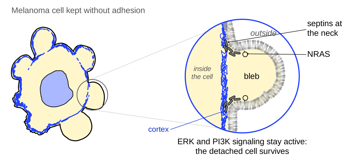
Blebs are usually discussed as protrusions of moving or dying cells, but a bleb also reshapes the membrane around it, and its narrow neck partly separates its contents from the rest of the cell. Both properties can matter for signaling. In melanoma cells kept without adhesion, the curved membrane at the edges of blebs, where each bleb meets the cell body, recruits septins, filament-forming proteins that bind curved membrane, and the septins scaffold an oncogenic form of the protein NRAS that keeps the ERK and PI3K survival pathways active (Weems et al., 2023). This septin signaling hub is strongly supported in cell lines in vitro, from one laboratory. The second half of the page turns to blebs as membrane compartments that concentrate some signaling lipids and proteins and exclude others.
Survival without adhesion
Most animal cells need attachment to survive. Without the signals that adhesion provides they undergo anoikis, a programmed cell death triggered by loss of attachment, and resisting anoikis is a key step for cancer cells that spread (Weems et al., 2023). Detached cells usually round up and bleb only for a while: blebbing fell from 62% to 10% of trypsin-detached CHO cells between 6 and 40 min (Kinn and Allen, 1981), and detached endothelial cells stopped blebbing within 40–60 min (Norman et al., 2010). Melanoma cells, in contrast, can keep blebbing indefinitely without attachment, and rounded, blebbing cells are common at the invasive front of tumors (Weems et al., 2023).
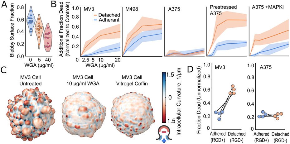
Weems et al. (2023) asked whether blebbing helps such cells survive, using MV3 melanoma cells, which carry an activating NRAS mutation. Wheat germ agglutinin (WGA), which reduces blebbing, killed more detached than adherent cells: after 24 h, the extra fraction of dead cells was about 0.5 for detached cells against about 0.17 for adherent ones (three paired experiments; read from plot). Enclosing single cells in bleb-restricting hydrogels gave the same result. A375 melanoma cells, which carry a BRAF mutation instead, were insensitive to WGA unless first stressed.
Curvature at bleb edges recruits septins
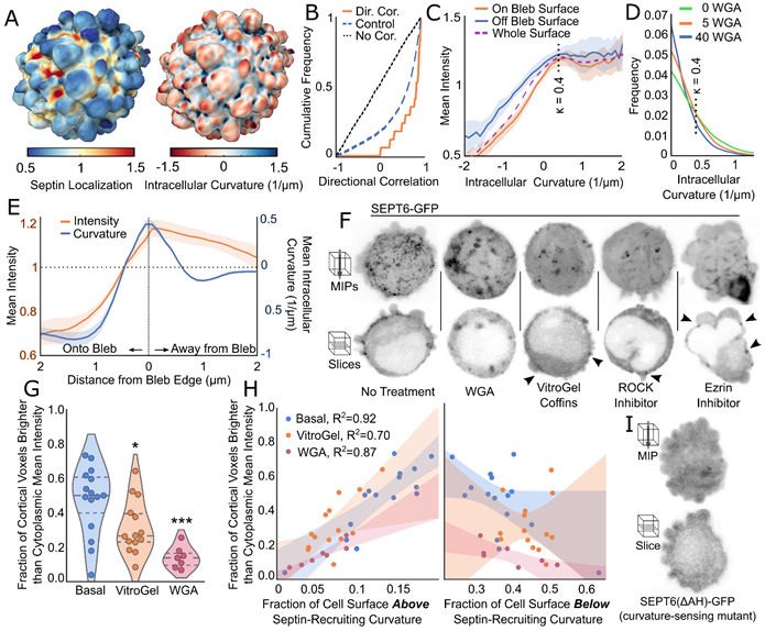
Weems et al. (2023) then mapped the curvature of the whole surface of MV3 cells in soft collagen. Seen from inside the cell, a bleb curves away (negative curvature), whereas the membrane at its edge, where it meets the cell body, bulges inward (positive curvature). A fluorescent septin, SEPT6, rose with positive curvature to a plateau from about 0.4 µm⁻¹, the curvature of a sphere 5 µm across (17 cells, 2,025 blebs; read from plot), and peaked at the edges of blebs.
All four treatments that reduced blebbing lowered cortical septin, although two were shown only as images, and in single cells cortical septin scaled with the fraction of the surface curved more tightly than 0.4 µm⁻¹ (R² = 0.70–0.92). A SEPT6 lacking its amphipathic helix, the segment that gives yeast septins their preference for curved membrane (Cannon et al., 2019), stayed off the membrane. The authors proposed that the septin layer integrates repeated blebbing while filtering out isolated blebs, an analysis that rests on a single cell (Weems et al., 2023).
From septins to survival signaling
What do the septins hold? Proximity labeling found NRAS among 321 septin partners, and NRAS lost its enrichment at the cell surface when septins were inhibited with the drug forchlorfenuron (FCF). Inhibiting either septins or blebs lowered the activity of ERK, a kinase downstream of NRAS, in detached but not in adherent cells, and lowered PI3K activity in rounded cells in collagen (Weems et al., 2023). The proposed chain therefore runs from blebs to curvature, to septins, to NRAS and to the ERK and PI3K pathways.
Each link was then tested for survival. FCF removed septin from the cell surface without changing blebbing and killed detached cells, and so did a dominant-negative NRAS. The most direct test used mouse embryonic fibroblasts, normal cells that stop blebbing soon after detachment. A dominant-negative dynamin kept them blebbing (about 80% at 3 h, against fewer than 10% of controls) and reduced the activation of caspases, the proteases that execute programmed cell death, and WGA or FCF reversed this protection. The authors concluded that sustained blebbing can make normal cells resist anoikis without any oncogenic mutation (Weems et al., 2023). The study has limits: septin and NRAS were overexpressed, the inhibitors may act on other targets, and many readouts rest on two to four experiments.
What curvature sensing by septins rests on
The evidence that septins read curvature comes mainly from reconstitution. Purified septin complexes bound lipid bilayers on silica beads best when the beads were 1–3 µm across (Bridges et al., 2016), and Cannon et al. (2019) traced this preference to the amphipathic helix. The value of 0.4 µm⁻¹ is less secure. None of the three in vitro studies that Weems et al. cite for it names a threshold, and in the third, septin density on wavy supported bilayers rose gradually to a maximum near +2 µm⁻¹, with no step at 0.4 µm⁻¹ (Beber et al., 2019). The cell data, too, show a graded rise rather than a switch. Geometry adds a caveat: on a rugged three-dimensional surface, even a uniform membrane label appears enriched near bleb edges (Driscoll et al., 2019), which Weems et al. answered in their main figures mainly with the helix mutant.
Independent support and conflicting results
One independent group has reported a related mechanism. Zhen et al. (2025) made detached MDA-MB-231 breast cancer cells bleb with latrunculin B, which depolymerizes actin, and found condensates of the adaptor protein SH3KBP1 that bridged Septin6 and a PI3K subunit at the bleb neck. Latrunculin lowered the death of detached cells, and WGA, FCF, a PI3K inhibitor or knockdown of SH3KBP1 each raised it again. Because the number of blebs was not measured, the link from blebs to survival in this system remains proposed.
Other results do not fit a single picture. In T lymphocytes, septins restrain blebbing: knocking down Septin 7 raised the fraction of blebbing cells from 9.9% to 29.7–37.1% (Tooley et al., 2009), and SEPT6 helped blebs retract (Gilden et al., 2012). In MV3 cells, by contrast, septin inhibition left blebbing unchanged (Weems et al., 2023).
Blebs as membrane compartments
A bleb can also hold signaling molecules apart from the rest of the cell surface, enriching some and depleting others. On the latrunculin-induced blebs of MDA-MB-231 cells, lipid sensors showed more PI(3,4,5)P3, the lipid that PI3K produces, and less of its substrate PI(4,5)P2 than on the rest of the membrane, while a general membrane marker was even (20 cells). A photoconverted PI(3,4,5)P3 sensor crossed the bleb neck poorly, and disrupting the septin–SH3KBP1 assembly at the neck abolished the difference (Zhen et al., 2025). Spontaneous blebs of MV3 cells instead carried more PI(4,5)P2 than the rest of the membrane (6 cells; Driscoll et al., 2019). Both studies used the same sensor, so in our reading the difference lies in how the blebs were made or in the cell type. Blebs may also confine damage, as discussed in Dying and injured cells (Babiychuk et al., 2011).
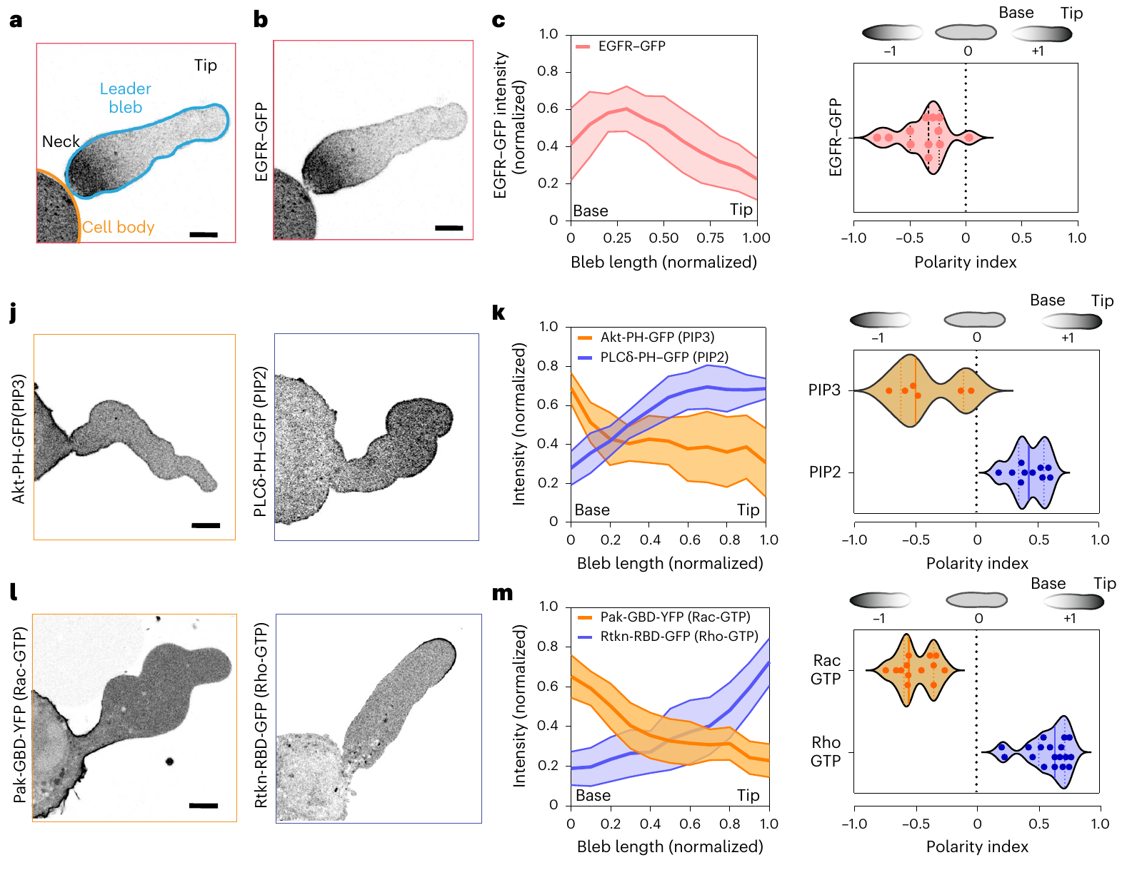
The neck matters most for the large leader blebs of confined cells. In confined A375 cells, ERK activity was enriched more than 250% in the leader bleb, against 10–12% for soluble proteins, and photoconverted protein left the bleb only slowly through its neck (Logue et al., 2015). Signaling is also graded along the leader bleb. In confined A375M2 melanoma cells, the EGF receptor, PI(3,4,5)P3 and active Rac were concentrated at the base of the bleb, and PI(4,5)P2 and active RhoA at its tip, the reverse of their order in crawling mesenchymal cells. The gradient depended on CD44, a transmembrane protein linked through ERM proteins to the dense actin meshwork at the bleb base, where it slowed the receptor. Depleting CD44 flattened the gradients of the receptor and of Rac activity and reduced migration led by leader blebs (Jha et al., 2026).
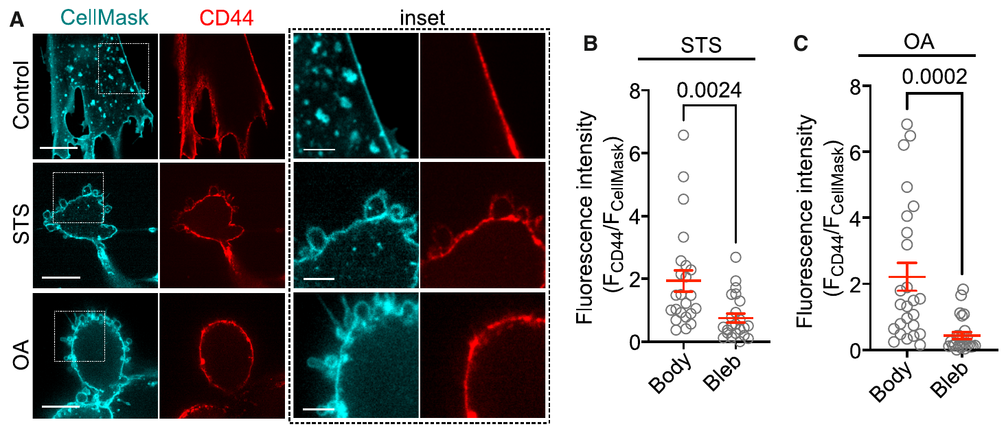
Blebs can also exclude proteins that the cortex holds. In fibroblasts made to bleb by apoptosis, or by okadaic acid, which makes the cortex contract without the lipid scrambling of apoptosis, the blebs lacked F-actin and membrane-bound ERM proteins. CD44, the receptor that carries the hyaluronan coat of the cell, and the transmembrane mucins were largely excluded from these blebs, and CD44 molecules inside a bleb diffused much faster (diffusion coefficient approaching 0.27 µm²/s, against about 0.03 µm²/s in the cell body). Without ERM proteins, or without its ERM-binding tail, CD44 entered the blebs. Apoptotic blebs therefore lacked the bulky hyaluronan coat and exposed phosphatidylserine, the “eat-me” lipid, to macrophages, whereas the untethered “don’t-eat-me” protein CD47 spread into them (Le et al., 2024).
In our reading, these studies suggest a common rule. A bleb membrane starts without a cortex, so molecules that diffuse freely, such as CD47 or lipid-anchored markers, spread evenly into it (Jha et al., 2026; Le et al., 2024). Transmembrane proteins tethered to cortical actin through ERM proteins, or corralled by such tethered proteins, stay where the cortex is dense: at the base of a leader bleb, or in the body of an apoptotic cell. Behind the neck, enzymes such as PI3K can then change the lipids locally. That blebs act as signaling compartments in this way, beyond the septin case, is proposed.
In tumors, and what remains open
The only test in tissue so far is a preprint (Weems et al., 2026 preprint). In samples from seven patients, mostly melanoma, rounded and blebbing cells showed septin enriched at the membrane (assessed qualitatively). In immunodeficient mice, oral FCF given after surgical removal of an MV3 tumor lowered recurrence from 66% to 23% (25 mice) and raised mean survival from 25.4 to 35.2 days (56 mice). Blebs were inferred from the shape of fixed cells, and only septins were perturbed, so FCF could act through septin functions unrelated to blebs. In our reading of the survival curves, treated mice began to die near the end of the 21-day treatment, which suggests that the drug delayed recurrence rather than prevented it.
Whether blebs themselves, rather than septins in general, support the survival of disseminating cells in living animals is therefore an open question; answering it needs a way to block blebbing that leaves septins intact. It is also unclear whether the signal is the curvature at bleb edges or the compartment behind a bleb neck, and whether other bleb-forming cells build such hubs.
The full evidence for the septin hub is on M16. How leader blebs move cells is described in Blebs in cell migration, and the fragments that blebs release in Fragments.
References (16)
- Babiychuk EB, Monastyrskaya K, Potez S et al. (2011) Blebbing confers resistance against cell lysis. Cell Death Differ 18:80–89. doi:10.1038/cdd.2010.81
- Beber A, Taveneau C, Nania M et al. (2019) Membrane reshaping by micrometric curvature sensitive septin filaments. Nat Commun 10:420. doi:10.1038/s41467-019-08344-5
- Bridges AA, Jentzsch MS, Oakes PW et al. (2016) Micron-scale plasma membrane curvature is recognized by the septin cytoskeleton. J Cell Biol 213:23–32. doi:10.1083/jcb.201512029
- Cannon KS, Woods BL, Crutchley JM et al. (2019) An amphipathic helix enables septins to sense micrometer-scale membrane curvature. J Cell Biol 218:1128–1137. doi:10.1083/jcb.201807211
- Driscoll MK, Welf ES, Jamieson AR et al. (2019) Robust and automated detection of subcellular morphological motifs in 3D microscopy images. Nat Methods 16:1037–1044. doi:10.1038/s41592-019-0539-z
- ◆ García-Arcos JM, Jha A, Waterman CM et al. (2024a) Blebology: principles of bleb-based migration. Trends Cell Biol 34:838–853. doi:10.1016/j.tcb.2024.02.009
- Gilden JK, Peck S, Chen YC et al. (2012) The septin cytoskeleton facilitates membrane retraction during motility and blebbing. J Cell Biol 196:103–114. doi:10.1083/jcb.201105127
- Jha A, Chandra A, Farahani PE et al. (2026) CD44 restricts EGFR mobility to polarize cytoskeletal signalling modules driving bleb-based migration. Nat Cell Biol 28:1408–1423. doi:10.1038/s41556-026-01981-1
- Kinn SR, Allen TD (1981) Conversion of blebs to microvilli: cell surface reorganisation after trypsin. Differentiation 20:168–173. doi:10.1111/j.1432-0436.1981.tb01172.x
- Le T, Ferling I, Qiu L et al. (2024) Redistribution of the glycocalyx exposes phagocytic determinants on apoptotic cells. Dev Cell 59:853–868. doi:10.1016/j.devcel.2024.01.020
- Logue JS, Cartagena-Rivera AX, Baird MA et al. (2015) Erk regulation of actin capping and bundling by Eps8 promotes cortex tension and leader bleb-based migration. eLife 4:e08314. doi:10.7554/eLife.08314
- Norman LL, Brugués J, Sengupta K et al. (2010) Cell blebbing and membrane area homeostasis in spreading and retracting cells. Biophys J 99:1726–1733. doi:10.1016/j.bpj.2010.07.031
- Tooley AJ, Gilden J, Jacobelli J et al. (2009) Amoeboid T lymphocytes require the septin cytoskeleton for cortical integrity and persistent motility. Nat Cell Biol 11:17–26. doi:10.1038/ncb1808
- Weems AD, Islam MT, Borges H et al. (2026) In vivo evidence for bleb-induced survival signaling in metastatic melanoma. bioRxiv. doi:10.64898/2026.01.05.697601 preprint
- Weems AD, Welf ES, Driscoll MK et al. (2023) Blebs promote cell survival by assembling oncogenic signalling hubs. Nature 615:517–525. doi:10.1038/s41586-023-05758-6
- Zhen Z, Zhang C, Li J et al. (2025) Blebs regulate phosphoinositide distribution and promote cell survival through the Septin-SH3KBP1-PI3K axis. Am J Physiol Cell Physiol 329:C145–C158. doi:10.1152/ajpcell.00096.2025